. 2013;8(1):e54519.
doi: 10.1371/journal.pone.0054519. Epub 2013 Jan 18.

Neural progenitor cell implants modulate vascular endothelial growth factor and brain-derived neurotrophic factor expression in rat axotomized neurons

Affiliations

Neural progenitor cell implants modulate vascular endothelial growth factor and brain-derived neurotrophic factor expression in rat axotomized neurons

Rocío Talaverón et al. PLoS One. 2013.

Abstract

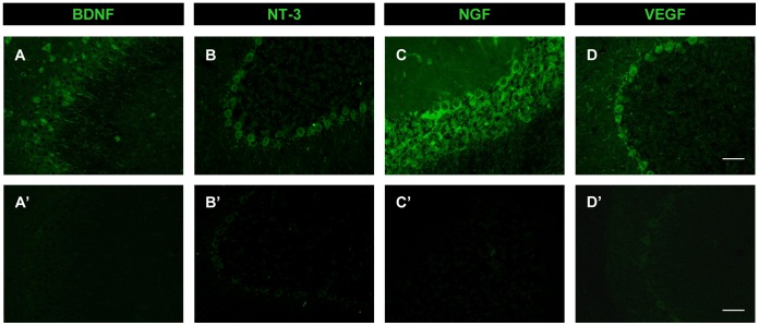
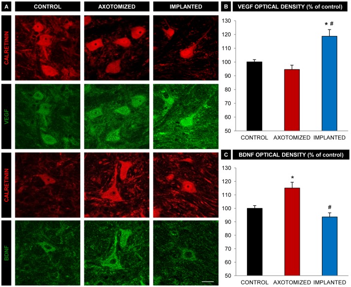
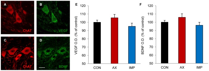
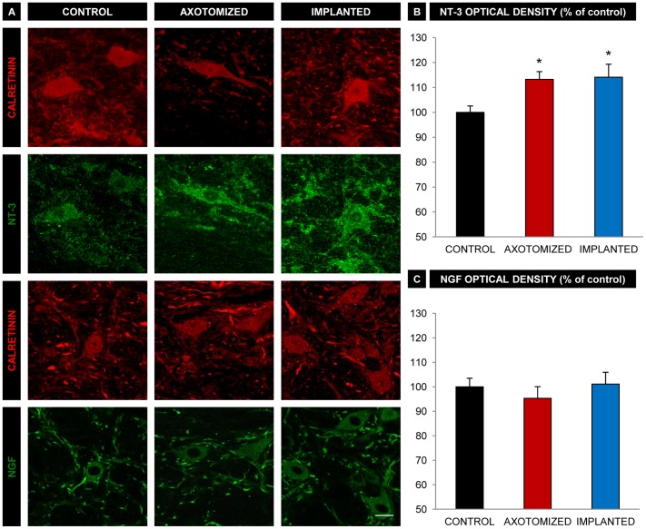
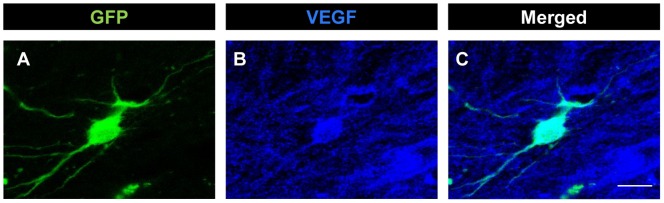
Axotomy of central neurons leads to functional and structural alterations which largely revert when neural progenitor cells (NPCs) are implanted in the lesion site. The new microenvironment created by NPCs in the host tissue might modulate in the damaged neurons the expression of a high variety of molecules with relevant roles in the repair mechanisms, including neurotrophic factors. In the present work, we aimed to analyze changes in neurotrophic factor expression in axotomized neurons induced by NPC implants. For this purpose, we performed immunofluorescence followed by confocal microscopy analysis for the detection of vascular endothelial growth factor (VEGF), brain-derived neurotrophic factor (BDNF), neurotrophin-3 (NT-3) and nerve growth factor (NGF) on brainstem sections from rats with axotomy of abducens internuclear neurons that received NPC implants (implanted group) or vehicle injections (axotomized group) in the lesion site. Control abducens internuclear neurons were strongly immunoreactive to VEGF and BDNF but showed a weak staining for NT-3 and NGF. Comparisons between groups revealed that lesioned neurons from animals that received NPC implants showed a significant increase in VEGF content with respect to animals receiving vehicle injections. However, the immunoreactivity for BDNF, which was increased in the axotomized group as compared to control, was not modified in the implanted group. The modifications induced by NPC implants on VEGF and BDNF content were specific for the population of axotomized abducens internuclear neurons since the neighboring abducens motoneurons were not affected. Similar levels of NT-3 and NGF immunolabeling were obtained in injured neurons from axotomized and implanted animals. Among all the analyzed neurotrophic factors, only VEGF was expressed by the implanted cells in the lesion site. Our results point to a role of NPC implants in the modulation of neurotrophic factor expression by lesioned central neurons, which might contribute to the restorative effects of these implants.


Conflict of interest statement

Competing Interests: The authors have declared that no competing interests exist.

Figures

Figure 1
Figure 1. Neural progenitor cell culture, axotomy and cell implant.
A. Floating neurosphere obtained from neural progenitors of the postnatal rat subventricular zone (SVZ). Scale bar: 25 µm. B. GFP-expressing cells (in green) in a SVZ-derived neurosphere. Scale bar: 25 µm. C. Schematic drawing of a rat parasagittal brainstem section showing the location of the medial longitudinal fascicle (MLF) transection and cell implant. Abducens internuclear neurons are represented in red and implanted neural progenitor cells in green. Axons of abducens internuclear neurons (red lines) course through the MLF towards the contralateral oculomotor nucleus. The distal stump of disrupted axons are represented in red dashed lines. D. Confocal microscopy image of a parasagittal brainstem section showing the implanted cells labeled with GFP (in green) at the site of axotomy. Dashed lines indicate the approximate dorso-ventral limits of the MLF. Scale bar: 100 µm. E. Higher magnification image of implanted GFP-labeled cells. Dorso-ventral and rostro-caudal orientation as in D. Scale bar: 50 µm. F. Schematic representation of a rat coronal section through the pons showing the abducens nucleus location. Abducens internuclear neuron somata are represented in red. Abbreviations: ABD: abducens nucleus; C: caudal; CB: cerebellum; D: dorsal; GFP: green fluorescent protein; MLF: medial longitudinal fascicle; OCM: oculomotor nucleus; py: pyramidal tract; R: rostral; V: ventral; 7n: facial nerve.
Figure 2
Figure 2. Antibody characterization.
Confocal microscopy images of rat coronal sections through the hippocampus (A, A’) and cerebellum (B, B’, C, C’) showing immunoreactivity to BDNF, NT-3, and NGF in positive controls (A, B and C, respectively) and in their respective blocking peptide-preadsorbed negative controls (A’, B’ and C’). D and D’. Confocal microscopy images of cerebellar coronal sections showing VEGF immunoreactivity in a positive control (D) and a negative control consisting of tissue pre-incubation with normal rabbit IgG instead of VEGF antibody (D’). Scale bars: 50 µm.
Figure 3
Figure 3. VEGF and BDNF immunoreactivity in abducens interneurons.
A. Confocal microscopy images of abducens internuclear neurons (immunopositive for calretinin, in red) showing double immunoreactivity against either VEGF (in green, second row of images) or BDNF (in green, fourth row). Examples of neurons from the different experimental conditions are shown: non lesioned neurons (CONTROL column), lesioned neurons from the axotomized group (AXOTOMIZED column) and lesioned neurons from the implanted group (IMPLANTED column). Bar: 20 µm. B. Optical density quantification of VEGF immunoreactivity in unlesioned neurons (CONTROL), lesioned neurons from the axotomized group (AXOTOMIZED) and lesioned neurons from the implanted group (IMPLANTED). Values are expressed as percentages relative to control (unlesioned neurons). Bars represent the mean ± SEM of 60–67 neurons analyzed from five different animals in each group. * and # indicate significant differences with respect to the control and the axotomized group, respectively (ANOVA test followed by Dunn’s method for multiple pairwise comparisons, p<0.05). C. Same as B, but for BDNF immunoreactivity. Bars represent the mean ± SEM of 32–54 neurons analyzed from four different animals in each group. * and # indicate significant differences with respect to the control and the axotomized group, respectively (ANOVA test followed by Dunn’s method for multiple pairwise comparisons, p<0.05).
Figure 4
Figure 4. VEGF and BDNF immunoreactivity in abducens motoneurons.
A to D. Confocal microscopy images of control abducens motoneurons showing double immunolabeling for ChAT (A, in red) and VEGF (B, in green), or for ChAT (C, in red) and BDNF (D, in green). Scale bar: 20 µm. E and F. Optical density (O.D.) quantification of VEGF immunoreactivity (E) and BDNF immunoreactivity (F) in abducens motoneurons from either the unlesioned side (CON), the lesioned side of the axotomized group (AX) or the lesioned side of the implanted group (IMP). Values represent percentages with respect to control (motoneurons from the unlesioned side). Bars show the mean ± SEM of 58–79 motoneurons analyzed from three different animals in each group. No significant differences were obtained between groups (ANOVA test followed by Dunn’s method for multiple pairwise comparisons).
Figure 5
Figure 5. NT-3 and NGF immunoreactivity in abducens interneurons.
A. Confocal images of abducens interneurons (immunopositive for calretinin, in red) showing double immunoreactivity against either NT-3 (in green, second row of images) or NGF (in green, fourth row). Examples of neurons from the different experimental conditions are shown: non lesioned neurons (CONTROL column), lesioned neurons from the axotomized group (AXOTOMIZED column) and lesioned neurons from the implanted group (IMPLANTED column). Scale bar: 20 µm B. Optical density quantification of NT-3 immunoreactivity in unlesioned neurons (CONTROL), lesioned neurons from the axotomized group (AXOTOMIZED) and lesioned neurons from the implanted group (IMPLANTED). Values are expressed as percentages with respect to control (unlesioned neurons). Bars represent the mean ± SEM of 25–38 neurons analyzed from four different animals in each group. *, p<0.05 compared to the control group, no significant differences were obtained between the axotomized and the implanted groups (ANOVA test followed by Dunn’s method for multiple pairwise comparisons, p<0.05). C. Same as B, but for NGF immunoreactivity. Bars represent the mean ± SEM of 26–54 neurons analyzed from three different animals in each group. Note that there were no significant differences between groups (ANOVA test followed by Dunn’s method for multiple pairwise comparisons, p<0.05).
Figure 6
Figure 6. VEGF immunoreactivity in neural progenitor implanted cells.
Confocal microscopy images of an implanted cell at the lesion site identified by its GFP expression (A, in green), which was also immunopositive for VEGF (B, in blue). C shows the merged image. Scale bar: 10 µm.

Similar articles

Cited by

References

    1. Titmus MJ, Faber DS (1990) Axotomy-induced alterations in the electrophysiological characteristics of neurons. Prog Neurobiol 35: 1–51. - PubMed
    1. Pastor AM, Delgado-García JM, Martínez-Guijarro FJ, López-García C, de la Cruz RR (2000) Response of abducens internuclear neurons to axotomy in the adult cat. J Comp Neurol 427: 370–390. - PubMed
    1. Giehl KM, Tetzlaff W (1996) BDNF and NT-3, but not NGF, prevent axotomy-induced death of rat corticospinal neurons in vivo. Eur J Neurosci 8: 1167–1175. - PubMed
    1. Kobayashi NR, Fan DP, Giehl KM, Bedard AM, Wiegand SJ, et al. (1997) BDNF and NT-4/5 prevent atrophy of rat rubrospinal neurons after cervical axotomy, stimulate GAP-43 and Talpha1-tubulin mRNA expression, and promote axonal regeneration. J Neurosci 17: 9583–9595. - PMC - PubMed
    1. Davis-López de Carrizosa MA, Morado-Díaz CJ, Tena JJ, Benítez-Temiño B, Pecero ML, et al. (2009) Complementary actions of BDNF and neurotrophin-3 on the firing patterns and synaptic composition of motoneurons. J Neurosci 29: 575–587. - PMC - PubMed

Publication types

Substances