. 2013;8(1):e54593.
doi: 10.1371/journal.pone.0054593. Epub 2013 Jan 17.

Sustained oxidative stress causes late acute renal failure via duplex regulation on p38 MAPK and Akt phosphorylation in severely burned rats

Affiliations

Sustained oxidative stress causes late acute renal failure via duplex regulation on p38 MAPK and Akt phosphorylation in severely burned rats

Yafei Feng et al. PLoS One. 2013.

Expression of concern in

Abstract

Background: Clinical evidence indicates that late acute renal failure (ARF) predicts high mortality in severely burned patients but the pathophysiology of late ARF remains undefined. This study was designed to test the hypothesis that sustained reactive oxygen species (ROS) induced late ARF in a severely burned rat model and to investigate the signaling mechanisms involved.

Materials and methods: Rats were exposed to 100°C bath for 15 s to induce severe burn injury (40% of total body surface area). Renal function, ROS generation, tubular necrosis and apoptosis, and phosphorylation of MAPK and Akt were measured during 72 hours after burn.

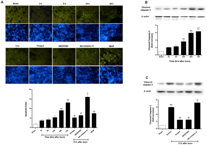
Results: Renal function as assessed by serum creatinine and blood urea nitrogen deteriorated significantly at 3 h after burn, alleviated at 6 h but worsened at 48 h and 72 h, indicating a late ARF was induced. Apoptotic cells and cleavage caspase-3 in the kidney went up slowly and turned into significant at 48 h and 72 h. Tubular cell ROS production shot up at 6 h and continuously rose during the 72-h experiment. Scavenging ROS with tempol markedly attenuated tubular apoptosis and renal dysfunction at 72 h after burn. Interestingly, renal p38 MAPK phosphorylation elevated in a time dependent manner whereas Akt phosphorylation increased during the first 24 h but decreased at 48 h after burn. The p38 MAPK specific inhibitor SB203580 alleviated whereas Akt inhibitor exacerbated burn-induced tubular apoptosis and renal dysfunction. Furthermore, tempol treatment exerted a duplex regulation through inhibiting p38 MAPK phosphorylation but further increasing Akt phosphorylation at 72 h postburn.

Conclusions: These results demonstrate that sustained renal ROS overproduction induces continuous tubular cell apoptosis and thus a late ARF at 72 h after burn in severely burned rats, which may result from ROS-mediated activation of p38 MAPK but a late inhibition of Akt phosphorylation.


Conflict of interest statement

Competing Interests: The authors have declared that no competing interests exist.

Figures

Figure 1
Figure 1. Blood urea nitrogen (BUN) and creatinine in rats after burn.
Analysis of burn-induced late renal dysfunction evidenced by BUN and creatinine (A–B) at different time points of 3 h, 6 h, 24 h, 48 h and 72 h and (C–D) with different treatments at 72 h postburn. The data showed that burn induced a late renal dysfunction at 72 h, which was improved by inhibiting reactive oxygen species (ROS) or p38 MAPK with tempol or SB203580, and deteriorated by inhibiting Akt expression at 72 h. Treatment with tBHP also caused significant renal dysfunction. *P<0.05, **P<0.01 vs. Sham; # P<0.05, ## P<0.01 vs. Vehicle. n = 6 rats per group per time point.
Figure 2
Figure 2. Histopathological renal injury in rats at 72 h after burn.
Representative images (magnification  = 200×) showed the histological examination for tubular damage with different treatments postburn using periodic acid–Schiff (PAS) staining. The data indicated burn induced significant renal damage at 72 h, which was improved by inhibiting ROS production or p38 MAPK expression, and deteriorated by inhibiting Akt expression. **P<0.01 vs. Sham; # P<0.05, ## P<0.01 vs. Vehicle. n = 6 rats per group per time point.
Figure 3
Figure 3. Tubular cell apoptosis and caspase-3 activation in rats after burn.
Burn induced (A) tubular apoptosis presented by renal TUNEL staining (magnification  = 400×) and increased caspase-3 activation (B) at different time points of 3 h, 6 h, 24 h, 48 h and 72 h (C) with different treatments at 72 h postburn. The data showed that burn induced increased caspase-3 activation and tubular cell apoptosis at a relatively late time stage postburn. Inhibition of ROS production and p38 MAPK phosphorylation alleviated renal apoptotic injury, while blockage of Akt expression aggravated the apoptosis in late renal failure after burn. Administration of tBHP led to obvious apoptotic kidney injury. **P<0.01 vs. Sham; # P<0.05, ## P<0.01 vs. Vehicle (72 h). n = 6 rats per group per time point.
Figure 4
Figure 4. Induction of oxidative stress in kidneys of rats postburn.
Burn injury induced sustained ROS generation evidenced by increased renal (A) H2O2 production, (B) thiobarbituric acid-reactive substances (TBARS) generation, suppressed antioxidant activity evidenced by decreased (C) SOD level and at 3 h, 6 h, 24 h, 48 h and 72 h after burn, which were attenuated by tempol at 72 h after burn. Renal (D) MnSOD activity and (E) protein expression, and (F) Nox4 expression was analyzed to determine the mitochondrial status. Treatment with tBHP also induced significant oxidative stress in kidneys of sham rats. *P<0.05, **P<0.01 vs. Sham; ## P<0.01 vs. Vehicle (72 h). n = 6 rats per group per time point.
Figure 5
Figure 5. Expression of MAPK and Akt signaling in kidneys of rats postburn.
Burn induced (A–C) elevated renal p38 MAPK phosphorylation in a time dependent manner but had no significant effect on ERK or JNK, whereas (D) Akt phosphorylation increased during the first 24 h but decreased beginning from 48 h after burn. **P<0.01 vs. Sham. n = 6 rats per group per time point.
Figure 6
Figure 6. Effect of ROS on expression of MAPK and Akt in kidneys of rats postburn.
(AB) Tempol attenuated ROS-induced phosphorylation of p38 and inhibition of Akt at 72 h postburn. (CD) Induction of ROS with tBHP significantly increased p38 but inhibited Akt phosphorylation. **P<0.01 vs. Sham; ## P<0.01 vs. Vehicle. n = 6 rats per group.
Figure 7
Figure 7. Schematic diagram showed the mediation of burn-induced sustained ROS overproduction on p38 MAPK and Akt expression and apoptotic pathway, which may be the mechanism of late acute renal failure.

Similar articles

Cited by

References

    1. Coca SG, Bauling P, Schifftner T, Howard CS, Teitelbaum I, et al. (2007) Contribution of acute kidney injury toward morbidity and mortality in burns: a contemporary analysis. Am J Kidney Dis 49: 517–523. - PubMed
    1. Mustonen KM, Vuola J (2008) Acute renal failure in intensive care burn patients (ARF in burn patients). J Burn Care Res 29: 227–237. - PubMed
    1. Chrysopoulo MT, Jeschke MG, Dziewulski P, Barrow RE, Herndon DN (1999) Acute renal dysfunction in severely burned adults. J Trauma 46: 141–144. - PubMed
    1. Holm C, Horbrand F, von Donnersmarck GH, Muhlbauer W (1999) Acute renal failure in severely burned patients. Burns 25: 171–178. - PubMed
    1. Mosier MJ, Pham TN, Klein MB, Gibran NS, Arnoldo BD, et al. (2010) Early acute kidney injury predicts progressive renal dysfunction and higher mortality in severely burned adults. J Burn Care Res 31: 83–92. - PMC - PubMed

Publication types

MeSH terms

Substances