. 2013;8(1):e54640.
doi: 10.1371/journal.pone.0054640. Epub 2013 Jan 18.

Mast cells express 11β-hydroxysteroid dehydrogenase type 1: a role in restraining mast cell degranulation

Affiliations

Mast cells express 11β-hydroxysteroid dehydrogenase type 1: a role in restraining mast cell degranulation

Agnes E Coutinho et al. PLoS One. 2013.

Abstract

Mast cells are key initiators of allergic, anaphylactic and inflammatory reactions, producing mediators that affect vascular permeability, angiogenesis and fibrosis. Glucocorticoid pharmacotherapy reduces mast cell number, maturation and activation but effects at physiological levels are unknown. Within cells, glucocorticoid concentration is modulated by the 11β-hydroxysteroid dehydrogenases (11β-HSDs). Here we show expression and activity of 11β-HSD1, but not 11β-HSD2, in mouse mast cells with 11β-HSD activity only in the keto-reductase direction, regenerating active glucocorticoids (cortisol, corticosterone) from inert substrates (cortisone, 11-dehydrocorticosterone). Mast cells from 11β-HSD1-deficient mice show ultrastructural evidence of increased activation, including piecemeal degranulation and have a reduced threshold for IgG immune complex-induced mast cell degranulation. Consistent with reduced intracellular glucocorticoid action in mast cells, levels of carboxypeptidase A3 mRNA, a glucocorticoid-inducible mast cell-specific transcript, are lower in peritoneal cells from 11β-HSD1-deficient than control mice. These findings suggest that 11β-HSD1-generated glucocorticoids may tonically restrain mast cell degranulation, potentially influencing allergic, anaphylactic and inflammatory responses.


Conflict of interest statement

Competing Interests: JRS holds patents on the use of 11beta-HSD1 inhibitors in diabetes, atherosclerotic disease and age-associated cognitive impairment and has been pursuing the development of 11beta-HSD1 inhibitors in an academic setting with submitted patents comprising composition of matter. There are no further patents, products in development or marketed products to declare. This does not alter the authors' adherence to all the PLOS ONE policies on sharing data and materials.

Figures

Figure 1
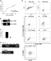
Figure 1. Mast cells express 11β-HSD1.
(A) 11β-HSD1 reductase (conversion of 11-dehydrocorticosterone to corticosterone; Solid lines/black squares) and dehydrogenase (conversion of corticosterone to 11-dehydrocorticosterone; dashed lines/open squares) activities in BMD-MC (>98% pure, assessed by tryptase staining as previously described [24]) are expressed as % conversion of 200 nM substrate by 2×10⁶ cells, over time (h). Values are mean ± SEM of 3 independent assays carried out on pooled BMD-MC. (B) BMD-MC express 11β-HSD1 mRNA (upper panel; 469 bp RT-PCR product with primers 868P and 869P) but not 11β-HSD2 mRNA (lower panel; 400 bp RT-PCR product). Lanes 5 and 6 show RT-PCR products from 2 independent Hsd11b1+/+ BMD-MC RNA samples. Positive controls (+) comprised liver mRNA (lane 4, upper panel) or kidney mRNA (lane 4, lower panel). Negative controls contained BMD-MC RNA but no reverse transcriptase (−, lane 3) or water (lane 2). Lane 1 contains a 100 bp ladder (Promega, Southampton, UK). (C) Hsd11b1−/− BMD-MC lack 11β-HSD1 activity. 11β-HSD1 reductase activity was measured in Hsd11b1+/+ (+/+) and Hsd11b1−/− (−/−) BMD-MC. Data are expressed as % conversion of 200 nM 11-dehydrocorticosterone to corticosterone by 2×10⁶ cells in a 10 h assay. Values are means of triplicate assays performed on pooled BMD-MC. ND, not detected. (D) Mast cells transcribe Hsd11b1 from the P1 promoter, whereas macrophages use the P2 promoter. Upper panel; RT-PCR products from 3 independent BMD-MC RNA samples showing the 627 bp RT-PCR product from P1 transcripts of 11β-HSD1 (lanes 4–6), but not P2 (predicted product of 647 bp, lanes 7–9) or P3 (predicted product of 542 bp, lanes 10–12). Kidney RNA confirmed transcription from P3 (not shown). Total 11β-HSD1 mRNA was detected using ex2 and 868P primers common to all transcripts (lanes 13–15, 587 bp product). Lower panel; RT-PCR products from BMD macrophage RNA showing the 647 bp RT-PCR P2 product (lane 3), but not the P1 (lane 2) or P3 products (lane 4). Total 11β-HSD1 mRNA (T) was detected using 868P and 869P primers, common to all transcripts (lanes 5, 6; 469 bp product). Lane 1 contains a 1 kb ladder (Invitrogen, Paisley, UK). Lanes marked (0) contain water only and lanes marked (−) show RT-PCR reactions from which the RT was omitted. (E) Flow cytometric staining revealed 11β-HSD1+CD117+ peritoneal cells. Freshly isolated peritoneal cells (∼5×10⁵ cells per sample) from male C57BL/6 mice were stained with 11β-HSD1 and CD117+ antibodies. Total peritoneal cells were first gated according to side scatter (SSC) and forward scatter (FSC) (i), then cells with high granularity, where mast cells lie (see Figure S1), were assessed for 11β-HSD1 and CD117 staining (ii–vii). Controls included; (ii) high SSC cells alone, (iii) sheep IgG control for 11β-HSD1 antibody, (iv) 11β-HSD1 antibody only, (v) isotype control for CD117 and (vi) CD117 antibody only. Panel (vii) shows cells double stained with 11β-HSD1 and CD117 antibodies.
Figure 2
Figure 2. More mast cells in the peritoneum of Hsd11b1−/− mice.
(A) Representative dot plots of freshly isolated peritoneal cells (5×10⁵) from Hsd11b1+/+ (left) and Hsd11b1−/− (middle) mice stained for CD117 (a mast cell marker) or isotype control (right) and subject to flow cytometry. (B) Peritoneal cells from Hsd11b1−/− mice (−/−, white bars) have more CD117+ cells than Hsd11b1+/+ (+/+, black bars), expressed as percentage of total cells (total cell number; Hsd11b1+/+, 3.8±0.5×10⁶ vs Hsd11b1−/−, 3.7±0.5×10⁶, p>0.05). (C) CD117+ cells from Hsd11b1−/− mice have higher mean fluorescence intensity (MFI) compared to Hsd11b1+/+ mice (black bars). Data are mean ± SEM, n = 8, *p<0.05, **p<0.01.
Figure 3
Figure 3. Decreased levels of glucocorticoid-sensitive transcripts in peritoneal cells from Hsd11b1−/− mice.
Real-time PCR measurement of (A) Carboxypeptidase A3 and (B) annexin 1 mRNA levels in total peritoneal cells from naïve male Hsd11b1−/− (−/−; white bars) or Hsd11b1+/+ (+/+; black bars) mice. Data are mean ± SEM; n = 8, *p<0.05, **p<0.01. CPA; carboxypeptidase A3.
Figure 4
Figure 4. Ultrastructural analysis suggests piecemeal degranulation in Hsd11b1−/− mast cells.
(A) Representative EM images showing peritoneal mast cells from Hsd11b1−/− (−/−) and Hsd11b1+/+ (+/+) mice. Closed arrowheads indicate dark (dense) granules and open arrowheads indicate lighter (less dense) granules. The lighter granules are suggestive of piecemeal degranulation in Hsd11b1−/− mice. MC; mast cell, G; granule, N; nucleus. (B) Peritoneal mast cells from Hsd11b1−/− (−/−) mice have fewer electron dense granules (black bars) and more “lighter” granules (white bars) compared to mast cells from Hsd11b1+/+ (+/+) mice. At least 40 individual mast cells from a pooled sample from each genotype were scored by 2 independent observers, blind to genotype. Data are expressed as mean ± SEM; **p<0.01.
Figure 5
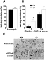
Figure 5. Hsd11b1−/− peritoneal mast cells are hyper-responsive to degranulation induced by K/BxN serum.
Release of β-hexosaminidase from peritoneal cells (2×10⁶) was measured following 15 min incubation with either (A) 10 µM ionomycin or (B) K/BxN serum (diluted 1∶8 or 1∶2 in Tyrode’s buffer). Black bars, Hsd11b1+/+; white bars, Hsd11b1−/−. Data are net degranulation in treated cells above levels measured in untreated cells (cells only are set to zero; see methods for details), and are mean ± SEM; n = 12−13, *p<0.05. (C) Representative micrographs (captured at 40× magnification) of enriched peritoneal CD117+ cells from Hsd11b1+/+ (+/+, left panels) or Hsd11b1−/− mice (−/−, right panels) following 21 h incubation with buffer (top panels) or K/BxN serum (1∶8 dilution) (lower panels). Arrowheads indicate degranulated mast cells.

Similar articles

Cited by

References

    1. Nigrovic PA, Lee DM (2005) Mast cells in inflammatory arthritis. Arthritis Res Ther 7: 1–11. - PMC - PubMed
    1. Kinet JP (2007) The essential role of mast cells in orchestrating inflammation. Immunol Rev 217: 5–7. - PubMed
    1. Laitinen LA, Laitinen A, Haahtela T (1993) Airway mucosal inflammation even in patients with newly diagnosed asthma. Am Rev Respir Dis 147: 697–704. - PubMed
    1. Pesci A, Foresi A, Bertorelli G, Chetta A, Olivieri D (1993) Histochemical characteristics and degranulation of mast cells in epithelium and lamina propria of bronchial biopsies from asthmatic and normal subjects. Am Rev Respir Dis 147: 684–689. - PubMed
    1. Nigrovic PA, Binstadt BA, Monach PA, Johnsen A, Gurish M, et al. (2007) Mast cells contribute to initiation of autoantibody-mediated arthritis via IL-1. Proc Natl Acad Sci U S A 104: 2325–2330. - PMC - PubMed

Publication types

MeSH terms

Substances