. 2013;8(1):e54758.
doi: 10.1371/journal.pone.0054758. Epub 2013 Jan 18.

The structure of the karrikin-insensitive protein (KAI2) in Arabidopsis thaliana

Affiliations

The structure of the karrikin-insensitive protein (KAI2) in Arabidopsis thaliana

Rohan Bythell-Douglas et al. PLoS One. 2013.

Abstract

KARRIKIN INSENSITIVE 2 (KAI2) is an α/β hydrolase involved in seed germination and seedling development. It is essential for plant responses to karrikins, a class of butenolide compounds derived from burnt plant material that are structurally similar to strigolactone plant hormones. The mechanistic basis for the function of KAI2 in plant development remains unclear. We have determined the crystal structure of Arabidopsis thaliana KAI2 in space groups P2(1) 2(1) 2(1) (a =63.57 Å, b =66.26 Å, c =78.25 Å) and P2(1) (a =50.20 Å, b =56.04 Å, c =52.43 Å, β =116.12°) to 1.55 and 2.11 Å respectively. The catalytic residues are positioned within a large hydrophobic pocket similar to that of DAD2, a protein required for strigolactone response in Petunia hybrida. KAI2 possesses a second solvent-accessible pocket, adjacent to the active site cavity, which offers the possibility of allosteric regulation. The structure of KAI2 is consistent with its designation as a serine hydrolase, as well as previous data implicating the protein in karrikin and strigolactone signalling.


Conflict of interest statement

Competing Interests: The authors have declared that no competing interests exist.

Figures

Figure 1
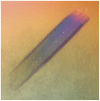
Figure 1. KAI2 crystal.
A multiple KAI2 crystal (32 μm×6 μm×6 μm) that was split for data collection (KAI2b).
Figure 2
Figure 2. Crystal structure of KAI2.
A. Stereoscopic ribbon diagram of KAI2 coloured from amino (blue) to carboxy (red) terminus. Every twentieth Cα is shown as a labelled sphere. B. Stereoscopic ribbon diagram of all three models of KAI2 (KAI2a blue, KAI2b orange, KAI2c black) and the models of DAD2 (purple) and RsbQ (brown) superposed. C. Stereoscopic cartoon diagram of KAI2. The α/β hydrolase domain is shown in blue and the cap domain shown in red.
Figure 3
Figure 3. Comparing KAI2 with DAD2.
The active site cavities of KAI2 (A) and of DAD2 (B). Cavity-exposed phenylalanine and tyrosine residues are shown for both proteins. Tyr124 of KAI2 occludes a small adjacent pocket that is not occluded in DAD2. Other than this difference, the two pockets are similar in size and shape. C. Sequence alignments of KAI2 and DAD2. The A. thaliana KAI2 sequence is coloured by similarity within the KAI2 protein family, the P. hybrida DAD2 sequence is coloured by similarity within the D14 protein family and the consensus sequence was determined and coloured by similarity across both families. Darker green colouration indicates more conserved sequence. Capital letters in the consensus sequence denote a conserved residue across all KAI2 and D14 proteins. Amino acids of interest are those where aligned KAI2 and D14 residues are coloured darker than the corresponding consensus sequence residue.
Figure 4
Figure 4. The KAI2 catalytic triad.
A. The catalytic residues Ser95-His246-Asp217 are hydrogen bonded in a classical arrangement for a serine hydrolase (Residues are shown in stick representation, coloured by atom type). B. CH…O hydrogen bonding between histidine Cε1 and carbonyl oxygens. KAI2 (blue), DAD2 (purple) and RsbQ (brown) all superimposed using the imidazole ring of the catalytic histidine of each respective protein. KAI2, unlike DAD2 and RsbQ, has unfavourable hydrogen bonding geometry between the catalytic His Cε1 and the carbonyl oxygen. This hydrogen bond is important for serine hydrolase activity , .
Figure 5
Figure 5. The second pocket of KAI2.
A. The two pockets of KAI2a are separated internally by the aromatic side-chain of Phe26. The active site residues can be seen in the primary pocket (left). Both pockets are solvent accessible. B. The active site cavity of DAD2 and the adjacent small, non-solvent accessible pocket.

References

    1. Flematti GR, Ghisalberti EL, Dixon KW, Trengove RD (2004) A compound from smoke that promotes seed germination. Science 305: 977. - PubMed
    1. Nelson DC, Flematti GR, Ghisalberti EL, Dixon KW, Smith SM (2012) Regulation of seed germination and seedling growth by chemical signals from burning vegetation. Annu Rev Plant Biol 63: 107–130. - PubMed
    1. Cook CE, Whichard LP, Turner B, Wall ME, Egley GH (1966) Germination of Witchweed (Striga lutea Lour.): Isolation and properties of a potent stimulant. Science 154: 1189–1190. - PubMed
    1. Cook CE, Whichard LP, Wall ME, Egley GH, Coggon P, et al. (1972) Germination Stimulants.II. Structure of strigol a potent seed-germination stimulant for witchweed (Striga-Lutea Lour). J Am Chem Soc 94: 6198–6199.
    1. Besserer A, Puech-Pagès V, Kiefer P, Gomez-Roldan V, Jauneau A, et al. (2006) Strigolactones stimulate arbuscular mycorrhizal fungi by activating mitochondria. PLoS Biol 4: e226. - PMC - PubMed

Publication types