. 2013;8(1):e54918.
doi: 10.1371/journal.pone.0054918. Epub 2013 Jan 22.

A comparison of Ku0063794, a dual mTORC1 and mTORC2 inhibitor, and temsirolimus in preclinical renal cell carcinoma models

Affiliations

A comparison of Ku0063794, a dual mTORC1 and mTORC2 inhibitor, and temsirolimus in preclinical renal cell carcinoma models

Hao Zhang et al. PLoS One. 2013.

Abstract

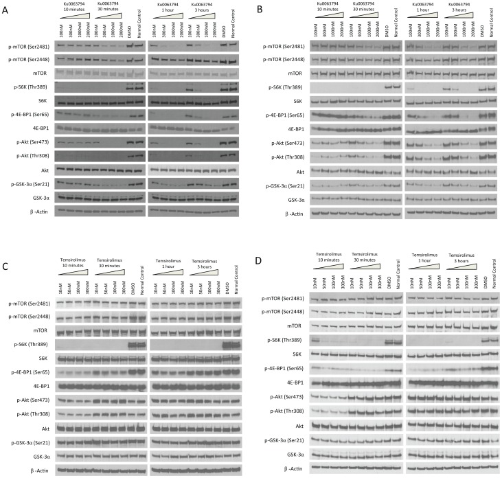
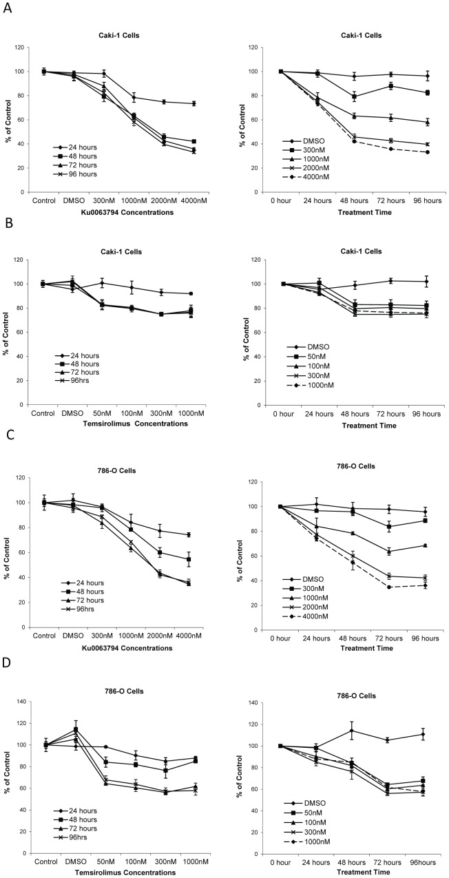
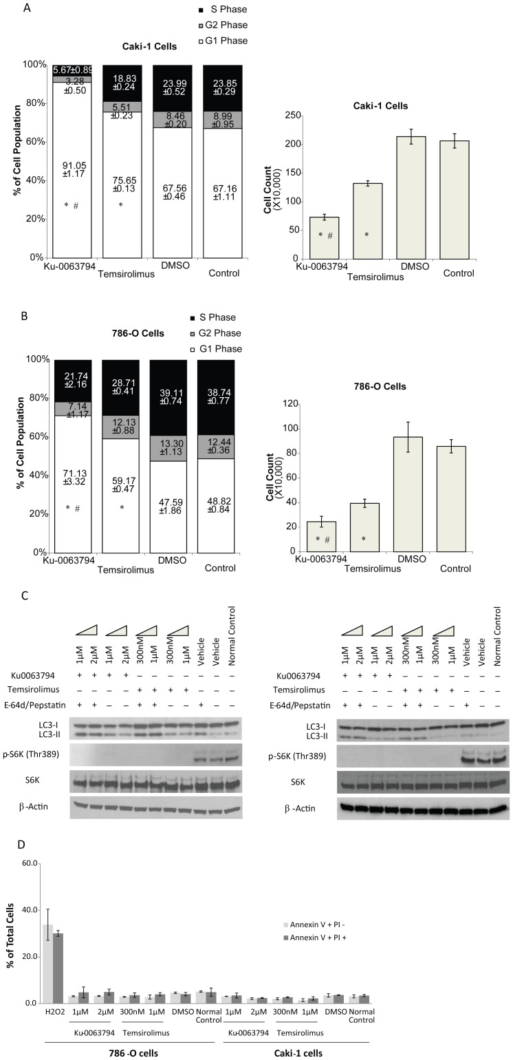
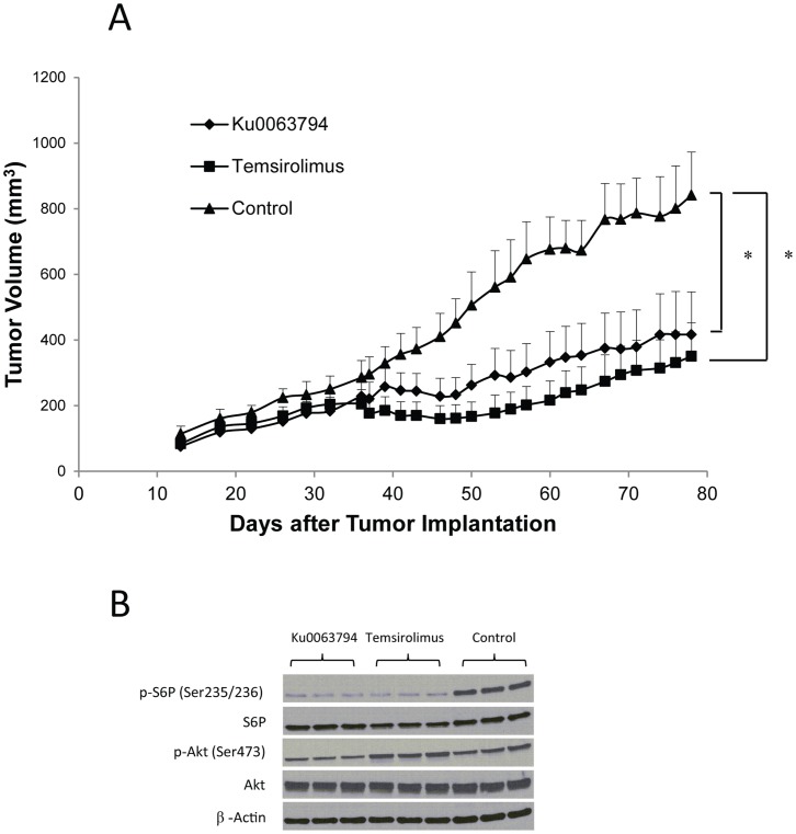
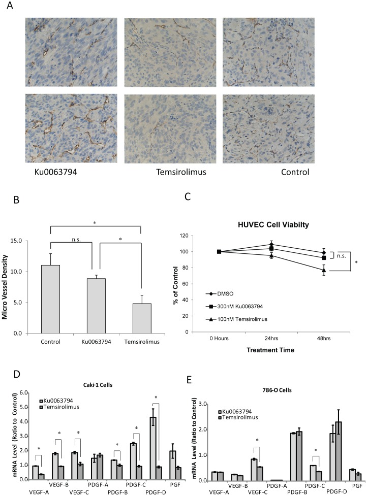
Rapamycin analogs, temsirolimus and everolimus, are approved for the treatment of advance renal cell carcinoma (RCC). Currently approved agents inhibit mechanistic target of rapamycin (mTOR) complex 1 (mTORC1). However, the mTOR kinase exists in two distinct multiprotein complexes, mTORC1 and mTORC2, and both complexes may be critical regulators of cell metabolism, growth and proliferation. Furthermore, it has been proposed that drug resistance develops due to compensatory activation of mTORC2 signaling during treatment with temsirolimus or everolimus. We evaluated Ku0063794, which is a small molecule that inhibits both mTOR complexes. Ku0063794 was compared to temsirolimus in preclinical models for renal cell carcinoma. Ku0063794 was effective in inhibiting the phosphorylation of signaling proteins downstream of both mTORC1 and mTORC2, including p70 S6K, 4E-BP1 and Akt. Ku0063794 was more effective than temsirolimus in decreasing the viability and growth of RCC cell lines, Caki-1 and 786-O, in vitro by inducing cell cycle arrest and autophagy, but not apoptosis. However, in a xenograft model there was no difference in the inhibition of tumor growth by Ku0063794 or temsirolimus. A potential explanation is that temsirolimus has additional effects on the tumor microenvironment. Consistent with this possibility, temsirolimus, but not Ku0063794, decreased tumor angiogenesis in vivo, and decreased the viability of HUVEC (Human Umbilical Vein Endothelial Cells) cells in vitro at pharmacologically relevant concentrations. Furthermore, expression levels of VEGF and PDGF were lower in Caki-1 and 786-O cells treated with temsirolimus than cells treated with Ku0063794.


Conflict of interest statement

Competing Interests: The authors have declared that no competing interests exist.

Figures

Figure 1
Figure 1. The mTOR pathway genes are overexpressed in ccRCC.
A heat map was generated with mTORC1 and mTORC2 genes that were significantly over-expressed in ccRCC when compared to normal kidney. Creighton examined genes induced by Akt using transgenic mice overexpressing AKT and identified 57 mTOR pathway genes that were positively correlated with AKT expression in human breast cancer (Oncogene, 26∶4648-55). The expression of these mTOR pathway genes, reported as rapalog sensitive (mTORC1) or insensitive (mTORC2), were examined in ccRCC.
Figure 2
Figure 2. Intracellular signaling in RCC cells treated with Ku0063794 or temsirolimus.
Caki-1 (A) and 786-O (B) cells were treated with Ku0063794 at the indicated concentrations for the indicated length of time. Control cells treated with DMSO were treated for 3 hrs. Cell lysates were used for western blotting to analyze the mTORC1/2 pathway. In an analogous experiment, Caki-1 (C) and 786-O (D) cells were similarly treated with temsirolimus or DMSO. The figure is representative of triplicate experiments.
Figure 3
Figure 3. Ku0063794 and temsirolimus decreased the viability of RCC cells.
Caki-1 (A, B) and 786-O (C, D) cells were plated on 96-well tissue-culture plates and then treated with Ku0063794 (A, C) or temsirolimus (B, D) at the indicated concentrations or DMSO (vehicle control) for 24 to 96 hours. Cell viability was calculated as a percent of the control and reported as a function of treatment time and drug concentration. The error bars indicate the standard error of the mean for experiments performed in triplicate.
Figure 4
Figure 4. Ku0063794 and temsirolimus induced G1 cell cycle arrest and autophagy in RCC cells.
Caki-1 (A) and 786-O (B) cells were plated and treated with 2 µM Ku0063794, 300 nM temsirolimus or DMSO (vehicle control) for 72 hours. The treated cells were subjected to cell cycle analysis (left). Cells were trypsinized and live cells were counted (right). The percent of cells at each cell cycle and the standard error of the mean are provided. *p<0.01 comparing either Ku0063794 or temsirolimus treatment to either DMSO treatment or untreated control. #p<0.01 comparing Ku0063794 to temsirolimus. Ku0063794 and temsirolimus induced autophagy in RCC cells as indicated by the increase in LC3-2/LC3-1 ratio. (C). Caki-1 (left) and 786-O (right) cells were treated with Ku0063794 or temsirolimus at the indicated concentration, or DMSO (vehicle control) for 24 hours with/without pepstatin A and E-64d. The figure is representative of triplicate experiments. (D) Ku0063794 and temsirolimus failed to induce apoptosis in RCC cells. Annexin-V and propidium iodide staining was performed following Ku0063794 or temsirolimus treatment at the indicated concentrations for 24 hours. As a positive control, 786-O cells were treated with 20 mM H2O2 for 30 minutes. Error bars indicate the standard error of the mean for triplicate experiments.
Figure 5
Figure 5. Ku0063794 and temsirolimus inhibited tumor growth in a xenograft model of RCC.
(A) The treatment with Ku0063794 or temsirolimus significantly inhibited tumor growth in Nu/Nu nude mice. Following subcutaneous injection of 786-O cells, mice (5 mice in each group) were treated from days 33–78. The error bars indicate the standard error of the mean. *p<0.05 when the Ku0063794 or temsirolimus group was compared with control. (B) 90 minutes after drug administration, both Ku0063794 and temsirolimus inhibited the mTORC1 pathway in vivo as indicated by decrease in S6P phosphorylation in the tumor tissues, while only Ku0063794 inhibited the mTORC2 pathway in vivo as indicated by decrease in Akt phosphorylation on Ser473. Each lane represents a tumor from a different mouse.
Figure 6
Figure 6. Temsirolimus but not Ku0063794 significantly inhibited tumor angiogenesis and viability of HUVEC cells at pharmacologically relevant concentrations.
(A) Following subcutaneous injection of 786-O cells, mice were treated with temsirolimus, Ku0063794 or control. The resulting tumors were stained with CD34 antibody and 2 representative images for each treatment group are shown. (B) The micro vessel density (MVD) of the tumors was quantified from at least 4 mice per group. The error bars indicate the standard error of the mean. *p<0.05 when the temsirolimus group was compared with Ku0063794 or control. n.s., not significant. (C) In vitro cell viability assay was performed using HUVEC cells treated with Ku0063794 or temsirolimus for 24 to 48 hours at pharmacologically relevant concentrations. The error bars indicate the standard error of the mean for experiments performed in triplicate. *p<0.01 when the temsirolimus group was compared with the DMSO group (control). (D, E) The mRNA levels of various angiogenic growth factors were measured using TaqMan® PCR in Caki-1 and 786-O cells before and after 24 hours of treatment with Ku0063794 (2 µM) or temsirolimus (300 nM). Error bars indicate the standard error of the mean for experiments performed in triplicate. *p<0.05 comparing Ku0063794-induced and temsirolimus-induced expression changes.

References

    1. Rini BI, Campbell SC, Escudier B (2009) Renal cell carcinoma. Lancet 373: 1119–1132. - PubMed
    1. Motzer RJ, Russo P (2000) Systemic therapy for renal cell carcinoma. J Urol 163: 408–417. - PubMed
    1. Furge KA, MacKeigan JP, Teh BT (2010) Kinase targets in renal-cell carcinomas: reassessing the old and discovering the new. Lancet Oncol 11: 571–578. - PubMed
    1. Foster KG, Fingar DC (2010) Mammalian target of rapamycin (mTOR): conducting the cellular signaling symphony. J Biol Chem 285: 14071–14077. - PMC - PubMed
    1. Zoncu R, Efeyan A, Sabatini DM (2011) mTOR: from growth signal integration to cancer, diabetes and ageing. Nat Rev Mol Cell Biol 12: 21–35. - PMC - PubMed

Publication types

MeSH terms

LinkOut - more resources