. 2013 Mar 15;437(2):81-8.
doi: 10.1016/j.virol.2012.11.013. Epub 2013 Jan 22.

IRES-based Venezuelan equine encephalitis vaccine candidate elicits protective immunity in mice

Affiliations

IRES-based Venezuelan equine encephalitis vaccine candidate elicits protective immunity in mice

Shannan L Rossi et al. Virology. .

Abstract

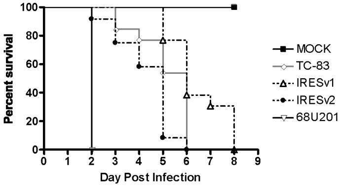
Venezuelan equine encephalitis virus (VEEV) is an arbovirus that causes periodic outbreaks that impact equine and human populations in the Americas. One of the VEEV subtypes located in Mexico and Central America (IE) has recently been recognized as an important cause of equine disease and death, and human exposure also appears to be widespread. Here, we describe the use of an Internal Ribosome Entry Site (IRES) from encephalomyocarditis virus to stably attenuate VEEV, creating a vaccine candidate independent of unstable point mutations. Mice infected with this virus produced antibodies and were protected against lethal VEEV challenge. This IRES-based vaccine was unable to establish productive infection in mosquito cell cultures or in intrathoracically injected Aedes taeniorhynchus, demonstrating that it cannot be transmitted from a vaccinee. These attenuation, efficacy and safety results justify further development for humans or equids of this new VEEV vaccine candidate.


Figures

Fig.1
Fig.1. Diagram of the wt 68U201 VEEV genome and IRES-based vaccine candidates
The mutated portion of the subgenomic promoter is shown on the top, with synonymous mutations shown in lowercase and stop codons highlighted in bold at the bottom.
Fig.2
Fig.2. In vitro characteristics of vaccine candidates, their parent strain 68U201, and vaccine strain TC-83
(A) Vero cell plaques measured 48 hpi in a 6-well plate. (B) Replication curves were performed in triplicate replicates at an MOI of 0.1 pfu/Vero cell. The timepoint at 1 hpi was below the limit of detection (dashed line) and arbitrarily assigned a titer of 1 pfu/ml. Error bars denote standard deviation.
Fig.3
Fig.3. Safety, immunogenicity and efficacy of vaccines in mice
Female CD1 mice, aged 6-8 weeks, were vaccinated or infected s.c. with 1×10⁵ pfu of virus, and weighed for 7-8 days thereafter. (A) The percent of initial weight following vaccination and (B) the percent of initial weight after challenge at 1, 3, or 12 months after vaccination. Error bars indicate standard deviation.
Fig.4
Fig.4. Viremia following vaccination
Sera taken on day 1 post vaccination were titrated for the presence of virus. The limit of detection is shown as a dashed line with a value of 67 pfu/ml. The combined data from three separate experiments are shown.
Fig.5
Fig.5. Immunogenicity of vaccines
Reciprocal PRNT80 titers at weeks 3, 6, 11 and 52 post vaccination were determined using VEEV strain 68U201 (IRES-based vaccines) or TC-83 (TC-83-vaccinated mice). Error bars denote standard error. All PRNT values <20 are recorded as 10, one dilution below the limit of detection. All PRNT titers >640 are reported as 640. The numbers above the bars denote the number of seropositive mice in each cohort.
Fig.6
Fig.6. Survival of 6-day-old mice injected intracranially with vaccines
Six-day-old CD1 mice were injected i.c. with 2×10⁴ pfu of each virus or MOCK-infected with PBS.
Fig.7
Fig.7. Infection of mosquito cells by vaccine strains
Vaccine stains and controls were passaged 5 times on C6/36 cells with a starting MOI of 0.1 Vero pfu/cell. (A) 48 hpi, supernatants were analyzed by RT-PCR with primers amplifying in the nsP4 region of the genome to detect the presence of viral RNA. (B) Titers obtained from each passage are shown. Error bars show standard deviation from duplicate samples.

References

    1. Adams AP, Navarro-Lopez R, Ramirez-Aguilar FJ, Lopez-Gonzalez I, Leal G, Flores-Mayorga JM, Rosa A.P.A.T.d., Saxton-Shaw KD, Singh AJ, Borland EM, Powers AM, Tesh RB, Weaver SC, Estrada-Franco JG. Venezuelan Equine Encephalitis Virus Activity in the Gulf Coast Region of Mexico, 2003-2010. PLoS Negl Trop Dis. 2012;6:e1875. - PMC - PubMed
    1. Brault AC, Powers AM, Holmes EC, Woelk CH, Weaver SC. Positively charged amino acid substitutions in the e2 envelope glycoprotein are associated with the emergence of venezuelan equine encephalitis virus. J Virol. 2002;76:1718–1730. - PMC - PubMed
    1. Brooke CB, Deming DJ, Whitmore AC, White LJ, Johnston RE. T cells facilitate recovery from Venezuelan equine encephalitis virus-induced encephalomyelitis in the absence of antibody. J Virol. 2010;84:4556–4568. - PMC - PubMed
    1. Burke DS, Ramsburg HH, Edelman R. Persistence in Humans of Antibody to Subtypes of Venezuelan Equine Encephalomyelitis (Vee) Virus after Immunization with Attenuated (Tc-83) Vee Virus-Vaccine. J Infect Dis. 1977;136:354–359. - PubMed
    1. Davis NL, Brown KW, Greenwald GF, Zajac AJ, Zacny VL, Smith JF, Johnston RE. Attenuated mutants of Venezuelan equine encephalitis virus containing lethal mutations in the PE2 cleavage signal combined with a second-site suppressor mutation in E1. Virology. 1995;212:102–110. - PubMed

Publication types

MeSH terms