. 2012 Dec 3;1(1):22.
doi: 10.1186/2046-2530-1-22.

Photoreceptor sensory cilia and ciliopathies: focus on CEP290, RPGR and their interacting proteins

Affiliations

Photoreceptor sensory cilia and ciliopathies: focus on CEP290, RPGR and their interacting proteins

Rivka A Rachel et al. Cilia. .

Abstract

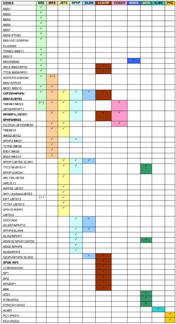
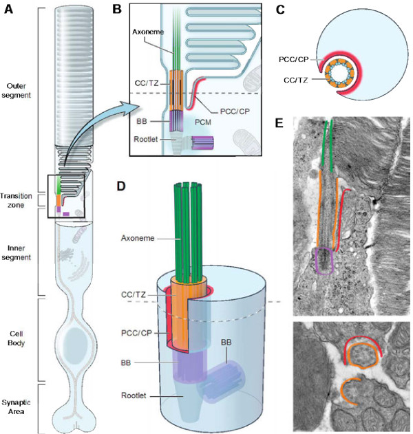
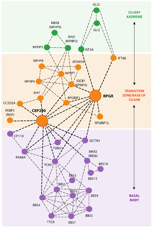
Ciliopathies encompass a broad array of clinical findings associated with genetic defects in biogenesis and/or function of the primary cilium, a ubiquitous organelle involved in the transduction of diverse biological signals. Degeneration or dysfunction of retinal photoreceptors is frequently observed in diverse ciliopathies. The sensory cilium in a photoreceptor elaborates into unique outer segment discs that provide extensive surface area for maximal photon capture and efficient visual transduction. The daily renewal of approximately 10% of outer segments requires a precise control of ciliary transport. Here, we review the ciliopathies with associated retinal degeneration, describe the distinctive structure of the photoreceptor cilium, and discuss mouse models that allow investigations into molecular mechanisms of cilia biogenesis and defects. We have specifically focused on two ciliary proteins - CEP290 and RPGR - that underlie photoreceptor degeneration and syndromic ciliopathies. Mouse models of CEP290 and RPGR disease, and of their multiple interacting partners, have helped unravel new functional insights into cell type-specific phenotypic defects in distinct ciliary proteins. Elucidation of multifaceted ciliary functions and associated protein complexes will require concerted efforts to assimilate diverse datasets from in vivo and in vitro studies. We therefore discuss a possible framework for investigating genetic networks associated with photoreceptor cilia biogenesis and pathology.


Figures

Figure 1
Figure 1
Ciliopathy genes with syndromic manifestations. Information from Online Mendelian Inheritance in Man (http://www.ncbi.nlm.nih.gov/omim). ALMS, Alstrom syndrome; BBS, Bardet–Biedl syndrome; COACH, Joubert syndrome with congenital hepatic fibrosis; JATD, Jeune asphyxiating thoracic dystrophy; JBTS, Joubert syndrome; LCA/RP, Leber congenital amaurosis/retinitis pigmentosa; MKKS, McKusick-Kaufman syndrome; MKS, Meckel–Gruber syndrome; NPHP, nephronophthisis; PKD, polycystic kidney disease; SLSN, Senior–Lø1ken syndrome.
Figure 2
Figure 2
Four distinct compartments in photoreceptor primary cilia, indicating known proteins that define their respective extent. The four compartments are: (1) distal cilium or axoneme (Axo; green); (2) connecting cilium/transition zone (CC/TZ; orange); (3) basal body (BB; purple); and (4) periciliary complex or ciliary pocket (PCC/CP; red). These compartments serve discrete functions in the cilium. (A) Schematic of a photoreceptor, showing specialized domains of the cell. The primary cilium elaborates into stacks of outer segment disks packed with rhodopsin, which serves as the primary light sensors of the cell. (B) Enlargement of the photoreceptor transition zone in two dimensions showing the four structural and functional domains in which most ciliary proteins are expressed. These domains are identified by known protein markers, such as acetylated α-tubulin (Axo + CC/TZ) and γ-tubulin (BB). Note: illustration of outer segment is based on a traditional model of disc morphogenesis in which nascent discs are open to the extracellular milieu, but a newer model posits that new discs form within the enclosure of outer segment plasma membrane [86]. (C) Cross-section through the CC/TZ of the photoreceptor showing the relationship between the microtubules of the cilium and the inner segment, via the PCC/CP. (D) Three-dimensional representation of the transition zone and adjacent domains shown in (B). Note the manner in which the PCC surrounds the TZ. Note also that the TZ is the one compartment that contacts all other compartments. (E) Electron micrographs showing longitudinal (top) and cross-section (bottom) views of mouse photoreceptors. Functional domains are highlighted with the corresponding colors shown in the other panels.
Figure 3
Figure 3
Interactome of ciliary proteins directly or indirectly connected to CEP290 and RPGR. Ciliary proteins directly (bold dotted lines) or indirectly (thin dotted lines) connected to CEP290 and RPGR. Ciliary expression domains are colored as in Figure 2. This network shows representative interactions.

Similar articles

Cited by

References

    1. Jain R, Pan J, Driscoll JA, Wisner JW, Huang T, Gunsten SP, You Y, Brody SL. Temporal relationship between primary and motile ciliogenesis in airway epithelial cells. Am J Respir Cell Mol Biol. 2010;43:731–739. doi: 10.1165/rcmb.2009-0328OC. - DOI - PMC - PubMed
    1. Sorokin SP. Reconstructions of centriole formation and ciliogenesis in mammalian lungs. J Cell Sci. 1968;3:207–230. - PubMed
    1. Berbari NF, O'Connor AK, Haycraft CJ, Yoder BK. The primary cilium as a complex signaling center. Curr Biol. 2009;19:R526–R535. doi: 10.1016/j.cub.2009.05.025. - DOI - PMC - PubMed
    1. Taschner M, Bhogaraju S, Lorentzen E. Architecture and function of IFT complex proteins in ciliogenesis. Differentiation. 2012;83:S12–S22. doi: 10.1016/j.diff.2011.11.001. - DOI - PMC - PubMed
    1. Wallingford JB, Mitchell B. Strange as it may seem: the many links between Wnt signaling, planar cell polarity, and cilia. Genes Dev. 2011;25:201–213. doi: 10.1101/gad.2008011. - DOI - PMC - PubMed

LinkOut - more resources