Review
. 2013:302:221-78.
doi: 10.1016/B978-0-12-407699-0.00004-2.

Beta-barrel scaffold of fluorescent proteins: folding, stability and role in chromophore formation

Affiliations
Review

Beta-barrel scaffold of fluorescent proteins: folding, stability and role in chromophore formation

Olesya V Stepanenko et al. Int Rev Cell Mol Biol. 2013.

Abstract

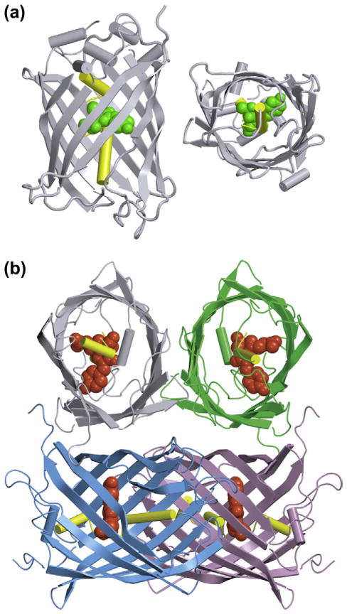
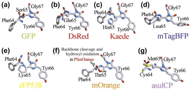
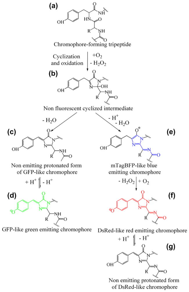
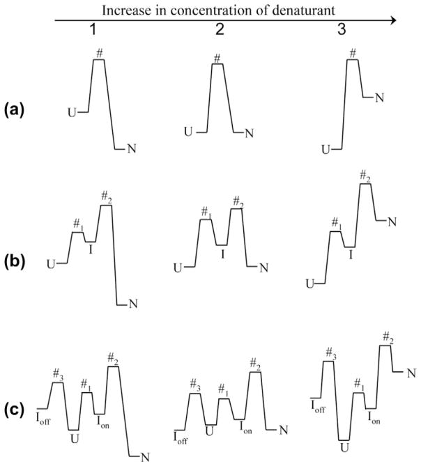
This review focuses on the current view of the interaction between the β-barrel scaffold of fluorescent proteins and their unique chromophore located in the internal helix. The chromophore originates from the polypeptide chain and its properties are influenced by the surrounding protein matrix of the β-barrel. On the other hand, it appears that a chromophore tightens the β-barrel scaffold and plays a crucial role in its stability. Furthermore, the presence of a mature chromophore causes hysteresis of protein unfolding and refolding. We survey studies measuring protein unfolding and refolding using traditional methods as well as new approaches, such as mechanical unfolding and reassembly of truncated fluorescent proteins. We also analyze models of fluorescent protein unfolding and refolding obtained through different approaches, and compare the results of protein folding in vitro to co-translational folding of a newly synthesized polypeptide chain.


Figures

Figure 4.1
Figure 4.1. Three-dimensional structure of sfGFP (PDB code 2B3P, Pedelacq et al., 2006) in two projections
(a) and of DsRed1 from Discosoma sp. (PDB code 1G7K, Yarbrough et al., 2001) (b). The chromophores of sfGFP and DsRed1 are shown as green and red space-filling unions, respectively. A central α-helix bearing the chromophore is shown in yellow. Monomers of DsRed1 are displayed in different colors. The drawing was generated by the graphic programs VMD (Humphrey et al., 1996) and Raster3D (Merritt and Bacon, 1997). (For interpretation of the references to color in this figure legend, the reader is referred to the online version of this book).
Figure 4.2
Figure 4.2. A variety of chromophore structures in FPs
a – green chromophore of GFP (PDB code 1W7S; van Thor et al., 2005); b and c – red chromophores of DsRed (PDB code 1G7K; Yarbrough et al., 2001) and Kaede (PDB code 2GW4; Hayashi et al., 2007); d – blue chromophore of mTagBFP (PDB code 3M24; Subach et al., 2010c); eg – derivatives of the DsRed-like red chromophore of zFP538 (PDB code 1XAE; Remington et al., 2005), mOrange (PDB code 2H5O; Shu et al., 2006), PSmOrange and asulCP (PDB code 2A50; Andresen et al., 2005). Carbon, nitrogen, oxygen and sulfur are colored in gray, blue, red and yellow, respectively. The drawing was generated based on the Protein Data Bank (Dutta et al., 2009) by the graphic programs VMD (Humphrey et al., 1996) and Raster3D (Merritt and Bacon, 1997). (For interpretation of the references to color in this figure legend, the reader is referred to the online version of this book).
Figure 4.3
Figure 4.3
General scheme of the autocatalytic synthesis of blue, green and red chromophores. (For interpretation of the references to color in this figure legend, the reader is referred to the online version of this book).
Figure 4.4
Figure 4.4. Change in the energy landscape induced by a denaturant
1 and 3 are the concentrations of denaturant at which the protein is in its native and unfolded states. U, N, I and # are the unfolded, native, intermediate and transition states of protein. Ion and Ioff are the on- and off-pathway intermediate states. a. Two-state unfolding–refolding model of protein. b. Three-state unfolding–refolding model of protein. c. Protein unfolding–refolding via on- and off-pathway intermediates.
Figure 4.5
Figure 4.5. sfGFP unfolding–refolding induced by GTC (Stepanenko et al., 2012b)
a, change in the absorption spectrum; b, chromophore fluorescence intensity corrected to the change of the chromophore absorption spectra and c, parameter A = I320/I356 of tryptophan fluorescence on GTC concentration. Inset to panel b: experimentally recorded chromophore fluorescence intensity (curve 1, gray), corrected to a total density of solution as follows I/W, where W = (1 − 10−DΣ)/DΣ (see Kuznetsova et al., 2012; Sulatskaya et al., 2011; Sulatskaya et al., 2012) (curve 2, pink), and corrected to the change of the chromophore absorption spectra (panel a) with the GTC concentration (curve 3, red). d, changes of the position of elution peaks of compact and denatured molecules (red and blue circles, respectively) and the change of the averaged elution volume of sfGFP (black triangles). Inset: Changes of the elution profile of sfGFP at increasing denaturant concentrations. The values of the curves specify applied denaturant concentration. (For interpretation of the references to color in this figure legend, the reader is referred to the online version of this book).
Figure 4.6
Figure 4.6. Structure of sfGFP
Localization and microenvironment of the chromophore and the tryptophan residue. a, diagram illustrating the formation of the β-barrel of 11 β-strands and the internal helix. The localization of the chromophore (Cro), tryptophan residue W57 and proline residues, including Pro89, the only proline that has the cis-conformation is shown; b, localization of α-helix in β-barrel. The β-barrel strand in the foreground is made transparent. The proline residues that are part of the α-helix, and Pro89, which is localized between the α-helix and the fourth β-strand, are shown; c, the microenvironment of W57; d, the chromophore microenvironment. (For interpretation of the references to color in this figure legend, the reader is referred to the online version of this book).
Figure 4.7
Figure 4.7. Reassembling sfGFP 1–10 and a synthetic 11-th β-strand
The truncated sfGFP 1–10 after refolding in native conditions does not reassemble with a synthetic peptide corresponding to 11-th β-strand, but it reassembles after light activation. The reassembled structure is identical to the native protein (Reprinted with permission from ( Kent and Boxer, 2011) Copyright 2011 American Chemical Society). (For color version of this figure, the reader is referred to the online version of this book).
Figure 4.8
Figure 4.8. Co-translational folding of FPs
a, a diagram of the FP fusion protein showing the C-terminal of the FP, the 6-amino acids linker and the N-terminal residues of the cystic fibrosis transmembrane conductance regulator (CFTR). The truncation sites are indicated by arrows. b, a diagram showing co-translational folding of the FP with different C-terminal tether lengths. c, the emission fluorescence spectra of the FP with different C-terminal tether lengths. d, the dependence of the intensity fluorescence of FP with different C-terminal tether lengths. (Adapted from figure 1 and figure 2 originally published in Journal of Biological Chemistry (Kelkar et al., 2012) Copyright 2012 the American Society for Biochemistry and Molecular Biology).

References

    1. Abbyad P, Childs W, Shi X, Boxer SG. Dynamic Stokes shift in green fluorescent protein variants. Proc Natl Acad Sci USA. 2007;104:20189–20194. - PMC - PubMed
    1. Adam V, Lelimousin M, Boehme S, Desfonds G, Nienhaus K, Field MJ, Wiedenmann J, McSweeney S, Nienhaus GU, Bourgeois D. Structural characterization of IrisFP, an optical highlighter undergoing multiple photo-induced transformations. Proc Natl Acad Sci USA. 2008;105:18343–18348. - PMC - PubMed
    1. Aglyamova GV, Hunt ME, Modi CK, Matz MV. Multi-colored homologs of the green fluorescent protein from hydromedusa Obelia sp. Photochem Photobiol Sci. 2011;10:1303–1309. - PubMed
    1. Ai HW, Henderson JN, Remington SJ, Campbell RE. Directed evolution of a monomeric, bright and photostable version of Clavularia cyan fluorescent protein: structural characterization and applications in fluorescence imaging. Biochem J. 2006;400:531–540. - PMC - PubMed
    1. Ai HW, Olenych SG, Wong P, Davidson MW, Campbell RE. Hue-shifted monomeric variants of Clavularia cyan fluorescent protein: identification of the molecular determinants of color and applications in fluorescence imaging. BMC Biol. 2008;6:13. - PMC - PubMed

Publication types

Substances