. 2013 Jan 24;20(1):82-91.
doi: 10.1016/j.chembiol.2012.11.005.

Effector kinase coupling enables high-throughput screens for direct HIV-1 Nef antagonists with antiretroviral activity

Affiliations

Effector kinase coupling enables high-throughput screens for direct HIV-1 Nef antagonists with antiretroviral activity

Lori A Emert-Sedlak et al. Chem Biol. .

Abstract

HIV-1 Nef, a critical AIDS progression factor, represents an important target protein for antiretroviral drug discovery. Because Nef lacks intrinsic enzymatic activity, we developed an assay that couples Nef to the activation of Hck, a Src family member and Nef effector protein. Using this assay, we screened a large, diverse chemical library and identified small molecules that block Nef-dependent Hck activity with low micromolar potency. Of these, a diphenylpyrazolo compound demonstrated submicromolar potency in HIV-1 replication assays against a broad range of primary Nef variants. This compound binds directly to Nef via a pocket formed by the Nef dimerization interface and disrupts Nef dimerization in cells. Coupling of nonenzymatic viral accessory factors to host cell effector proteins amenable to high-throughput screening may represent a general strategy for the discovery of new antimicrobial agents.


Figures

Figure 1
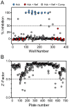
Figure 1. High-throughput screening for inhibitors of Nef-dependent Hck activity
The NIH Molecular Libraries Screening Centers Network (MLSCN) library (~220,000 compounds) was screened using the FRET-based Nef:Hck in vitro kinase assay as described in the text. (a) Scatterplot of results from a representative 384-well plate. Under these conditions, Hck is inactive when added by itself (blue circles), while addition of a 10-fold molar excess of Nef induces Hck kinase activation, demonstrating the Nef dependence of the assay (red circles). Compounds were screened at 20 μM under conditions where Hck activity is completely dependent on Nef (grey circles), with > 50% inhibition defined as a ‘hit’ (dashed line). (b) Z′-factors for each 384-well plate for the entire high-throughput screening campaign. Of 694 plates screened, 684 passed with Z′-factors ≥ 0.5 (98.5% pass rate); plates that failed due to robotic error were rescreened and subsequently passed (see also Table S1).
Figure 2
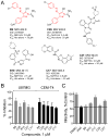
Figure 2. Inhibitors of Nef-dependent Hck activity also block Nef-dependent HIV replication and infectivity
(a) Chemical structures of the compounds identified by the Nef:Hck HTS campaign and subsequently shown to block Nef-dependent enhancement of HIV-1 replication in two different cell lines are shown, along with the IC50 values for each compound against Hck alone vs. the Nef:Hck complex. Two compounds from the screen share a diphenyldiazenylpyrazole substructure (highlighted in red), differing only in the placement of a single chlorine atom. (b) HIV replication. Compounds (1 μM) shown in (a) were added to cultures of U87MG and CEM-T4 cells in 96-well plates, followed by infection with wild-type HIV-1 NL4-3 (50 pg p24 equivalents/ml) 1 h later. Viral output for the DMSO-treated control cultures was consistently greater than 100-fold over the HIV input, typically ranging from 9,000–12,000 p24 equivalents/ml. Virus replication was assessed by p24 ELISA after 4 days (U87MG) or 9 days (CEM-T4). Data are expressed as the mean percent inhibition as compared to control cultures incubated with the carrier solvent (DMSO) ± S.E.M. (n=4). Results of replication and cytotoxicity experiments with all 66 hit compounds from the primary screen are shown in Figures S1 and S2. (c) Infectivity assays. Compounds (3 μM) were added to cultures of the reporter cell line TZM-bl followed by infection with either wild-type or Nef-defective (ΔNef) HIV NL4-3 in 96-well plates. After 48 hours, relative virus infectivity was assessed as luciferase production in infected cells. Results are plotted as the mean percent of HIV-1 infectivity observed in control cells incubated with the carrier solvent DMSO ± S.E.M. (n=3). In the absence of Nef, infectivity is reduced by about 50% (ΔNef; dashed line shown for reference). See also Figures S1 and S2.
Figure 3
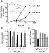
Figure 3. Inhibition of Nef-dependent Hck activity and HIV-1 replication by the diphenylpyrazolo compound, B9
(a) Concentration-response curves for B9 were generated with the Nef:Hck complex (circles) vs. Hck alone (squares) using the Z′-lyte kinase assay. For Hck alone, approximately 5-fold more kinase protein was added to achieve a similar level of activity as the Nef:Hck complex. Under these conditions, B9 inhibits Nef-dependent Hck activation with an IC50 value in the low micromolar range (2.8 μM), while the IC50 value for Hck alone is > 20 μM. Kinase assays were performed three times in quadruplicate, and the data represent percent inhibition as compared to the DMSO vehicle control ± S.E.M. (b) CEM-T4 cells were infected with wild-type HIV-1 NL4-3 (grey bars) or the corresponding Nef-defective mutant (ΔNef; black bars) in the presence of the B9 concentrations shown. Viral replication was assessed 9 days later by p24 ELISA. Input virus for HIV-1 ΔNef was increased by ten-fold relative to wild-type to compensate for the reduced infectivity and replication of Nef-defective virus in CEM-T4 cells (Narute and Smithgall, 2012). This experiment was done in triplicate and data are represented as percent of HIV-1 replication relative to the DMSO vehicle control ± S.E.M. (c) TZM-bl cells were infected with wild-type (gray bars) and ΔNef (black bar) HIV NL4-3 in the presence of the B9 concentrations shown, and infectivity was assessed as luciferase activity 48 h later. This experiment was repeated three times in triplicate and the data are represented as percent infectivity relative to the DMSO control ± S.E.M. In the absence of Nef, infectivity is reduced by about 50% (dashed line shown for reference).
Figure 4
Figure 4. Inhibition of HIV-1 Nef chimera replication and endogenous SFK activation in CEM-T4 cells by the diphenylpyrazolo compound, B9
(a) CEM-T4 cells (1 × 10⁴ per well of a 96-well plate) were infected with wild-type HIV-1 NL4-3, a Nef-defective mutant (ΔNef), or the indicated Nef chimeras in a final culture volume of 200 μl. Input virus for HIV-1 ΔNef was increased by ten-fold relative to wild-type to compensate for the reduced infectivity and replication of Nef-defective virus in CEM-T4 cells (Narute and Smithgall, 2012). B9 was added to the cultures to final concentrations of 0.3 and 1.0 μM, and viral replication was determined by p24 ELISA 10 days later. Data are expressed as the mean percent of HIV-1 replication observed in control cultures incubated with the carrier solvent (0.1 % DMSO) ± S.D. (n=6). (b) CEM-T4 cells were infected with wild-type HIV-1 NL4-3, a Nef-defective mutant (ΔNef), or the indicated Nef chimeras in a final culture volume of 10 ml in the presence of B9 (1 μM) or the DMSO carrier solvent as a control (Con). The infected cells were lysed and Src-family kinase proteins were immunoprecipitated with a pan-specific antibody and protein G-sepharose beads. The SFK activation state was assessed by immunoblotting with a phosphospecific antibody against the activation loop phosphotyrosine residue common to all Src-family members (pY418). Control blots were performed on cell lysates for HIV-1 Gag proteins (p55, p40, and p24), Nef, as well as actin as a loading control. Results from uninfected cells are shown in the far right lane (No virus). This experiment was repeated twice with comparable results.
Figure 5
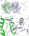
Figure 5. Docking studies predict direct interaction of B9 with the HIV-1 Nef dimer interface
(a) The two “halves” of the Nef dimer (PDB: 1EFN) are modeled in green and blue, respectively (Nef-A and Nef-B). B9 is docked at the two most energetically favored binding sites (Sites 1 and 2). The structure of B9 is shown for reference (right). (b) Close-up view of the predicted B9 binding sites. Site 1 is more energetically favored and nestles between the α-helices that form the dimer interface. Here B9 is predicted to form an extensive network of polar contacts with Nef residues Gln104, Gln107, and Asn126. Site 2 is positioned on the surface of each Nef monomer, away from the dimerization interface, and also makes a polar contact with Asn126. A single site 2 interaction of B9 with the Nef dimer is shown for simplicity. B9 also docks to the SIV Nef dimerization interface; see Figure S3. For additional details of docking results, see Table S3.
Figure 6
Figure 6. B9 binding to Nef requires the predicted binding pocket residue Asn126
(a) Surface Plasmon Resonance. Recombinant purified HIV-1 Nef-SF2 was immobilized on the surface of a Biacore CM5 chip and B9 was flowed past Nef at the concentrations shown. The flow path was switched back to buffer after 180 s to induce B9 dissociation (arrow). The resulting sensorgrams (black lines) were best-fit by a heterogeneous ligand (Nef in this case) model (red lines) supporting the presence of two distinct binding sites with Kd values of 860 ± 58 nM and 1.72 ± 0.23 nM. (b) SPR data were also fit by a two-state model, which yielded a Kd value 1.79 ± 0.11 nM for the final Nef:B9 complex. (c) SPR analysis was repeated with wild-type (WT) Nef and three Nef mutants in which Asn126 is replaced with Leu, Gln, or Ala as shown. B9 was held constant at 10 μM and bound readily to wild-type Nef but not to any of the N126 mutants. (d) Nef Asn126 mutants retain their ability to activate Hck. Downregulated Hck was assayed in vitro using the Z′-Lyte kinase assay and Tyr2 peptide substrate either alone or in the presence of a 10-fold molar excess of wild-type Nef or the three Asn126 mutants shown. All four Nef proteins produced an equivalent shift of the Hck activation curve to the left, indicating that mutagenesis of Asn126 does not affect its ability to bind and activate Hck (see also Table S3).
Figure 7
Figure 7. B9 inhibits Nef dimerization in cells
A) Molecular model of the Nef dimerization interface, based on the crystal structure of the Nef:SH3 complex (Lee et al., 1996). Hydrophobic side chains that contribute to dimerization are indicated; substitution of these residues with aspartate (Nef-4D mutant) dramatically reduces Nef dimerization as determined by fluorescence complementation assay (Poe and Smithgall, 2009). B) Human 293T cells were transfected with Nef-BiFC constructs and incubated with B9 over a range of concentrations. The Nef-4D mutant was included as a negative control. Following incubation for 48 hours, the cells were fixed, stained with a Nef antibody and Texas-red, and analyzed by two-color fluorescence microscopy. The top panel shows representative images from cells expressing the wild-type Nef (Nef-WT) in the absence or presence of B9 (6 μM) as well as the dimerization defective mutant, Nef-4D. The bottom panel shows the result of image analysis, in which BiFC (Nef dimerization) to immunofluorescence (Nef expression) intensity ratios were calculated for at least 150 cells. Data were normalized to the untreated Nef-WT control, and represent the mean ± S.D. C) Nef-4D fails to activate Hck. Downregulated Hck was assayed in vitro using the Z′-Lyte kinase assay and Tyr2 peptide substrate either alone or in the presence of a 10-fold molar excess of either wild-type Nef (Nef-WT) or the Nef-4D mutant.

Similar articles

Cited by

References

    1. Aiken C, Trono D. Nef stimulates human immunodeficiency virus type 1 proviral DNA synthesis. J Virol. 1995;69:5048–5056. - PMC - PubMed
    1. Arold ST, Baur AS. Dynamic Nef and Nef dynamics: how structure could explain the complex activities of this small HIV protein. Trends Biochem Sci. 2001;26:356–363. - PubMed
    1. Atkins KM, Thomas L, Youker RT, Harriff MJ, Pissani F, You H, Thomas G. HIV-1 Nef binds PACS-2 to assemble a multikinase cascade that triggers major histocompatibility complex class I (MHC-I) down-regulation: analysis using short interfering RNA and knock-out mice. J Biol Chem. 2008;283:11772–11784. - PMC - PubMed
    1. Betzi S, Restouin A, Opi S, Arold ST, Parrot I, Guerlesquin F, Morelli X, Collette Y. Protein protein interaction inhibition (2P2I) combining high throughput and virtual screening: Application to the HIV-1 Nef protein. Proc Natl Acad Sci U S A. 2007;104:19256–19261. - PMC - PubMed
    1. Chowers MY, Spina CA, Kwoh TJ, Fitch NJ, Richman DD, Guatelli JC. Optimal infectivity in vitro of human immunodeficiency virus type 1 requires an intact nef gene. J Virol. 1994;68:2906–2914. - PMC - PubMed

Publication types

MeSH terms

Substances

LinkOut - more resources