. 2013 Jan 24;20(1):102-10.
doi: 10.1016/j.chembiol.2012.10.021.

Biofilm inhibitors that target amyloid proteins

Affiliations

Biofilm inhibitors that target amyloid proteins

Diego Romero et al. Chem Biol. .

Abstract

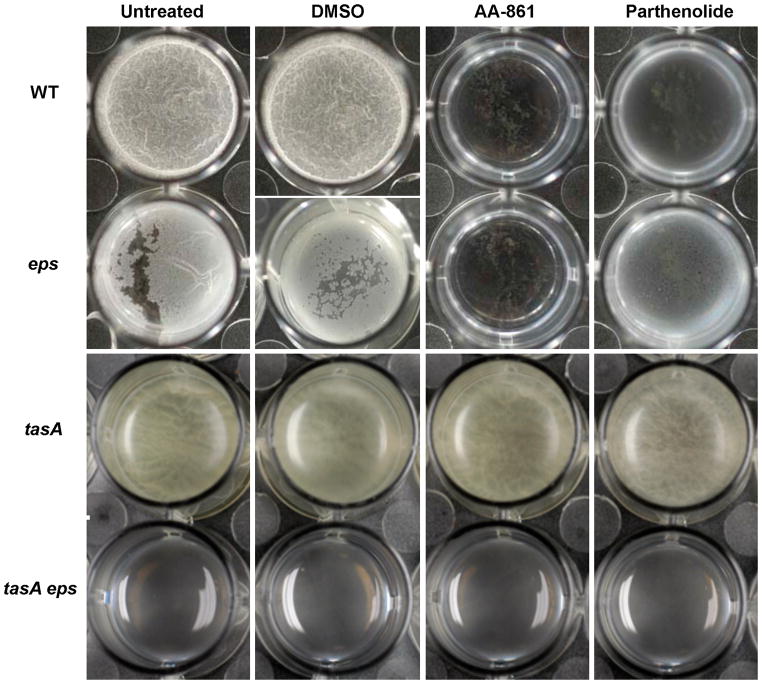
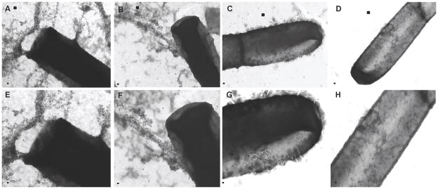
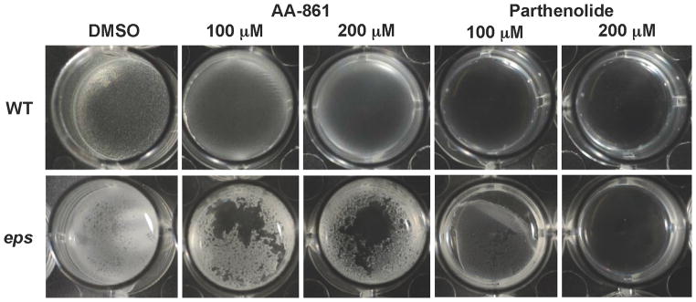
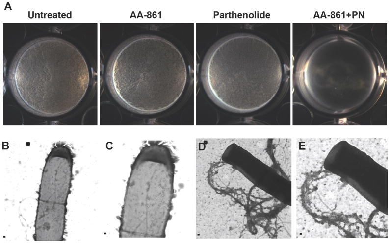
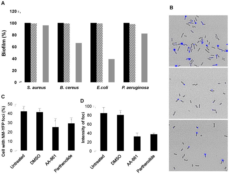
Bacteria establish stable communities, known as biofilms, that are resistant to antimicrobials. Biofilm robustness is due to the presence of an extracellular matrix, which for several species-among them Bacillus subtilis-includes amyloid-like protein fibers. In this work, we show that B. subtilis biofilms can be a simple and reliable tool for screening of molecules with antiamyloid activity. We identified two molecules, AA-861 and parthenolide, which efficiently inhibited biofilms by preventing the formation of amyloid-like fibers. Parthenolide also disrupted pre-established biofilms. These molecules also impeded the formation of biofilms of other bacterial species that secrete amyloid proteins, such as Bacillus cereus and Escherichia coli. Furthermore, the identified molecules decreased the conversion of the yeast protein New1 to the prion state in a heterologous host, indicating the broad range of activity of the molecules.


Figures

Figure 1
Figure 1. Screening of molecules with anti-biofilm activity
384 well microplates filled with MSgg medium were inoculated with B. subitlis 3610 cells and aliquots of a collection of small molecules at a final concentration of 12.5 μg/ml were added. After 24 h of incubation, plates were assessed for presence or absence of pellicles. (A) A close view of one of the plates showing the inhibition of pellicle provoked by two different molecules, (B) Structure of AA-861, a benzoquinone derivative, and (C) parthenolide, a sesquiterpene lactone. (D) A growth curve of B. subtilis 3610 in MSgg liquid medium showed no variation in bacterial growth in the absence (○) or presence of 50 μM of AA-861 (■), or parthenolide (×).
Figure 2
Figure 2. TasA is the main target of the anti-biofilm molecules
Biofilm assays were performed to determine the main target of AA-861 and parthenolide. MSgg medium was inoculated with cells of wild-type, eps or tasA single mutants or double tasA, eps mutant. The molecules were added at 50 μM. DMSO was used as a negative control. Images of pellicles were taken after 24 hr of incubation at 30°C with no agitation (see also Figure S1 and Figure S2).
Figure 3
Figure 3. The molecules inhibit fibers formation in cell surfaces
Transmission electron micrographs of negatively stained, anti-TasA immunogold labeled samples from pellicles grown for 24 hr in MSgg medium. (A) Untreated cells or (B) cells treated with DMSO as a negative control show wild-type fibers. Cells treated with 50 μM of (C) AA-861 and (D) parthenolide did not show fibers although the gold-labeled antibodies remained associated to cell surfaces. Panels E–H represent a magnified view of areas marked in panels A–D. Scale bars, 100 nm in panels A–D and 200 nm in panels E–H (see also Figure S2).
Figure 4
Figure 4. The molecules inhibit the polymerization of TasA in vitro
(A) Thioflavin T and fluorescence were used to study the kinetics of polymerization of TasA. Purified TasA was treated with formic acid, dried under vacuum and mixed with physiological buffer (○) or DMSO (▲), plus 50 μM of AA-861 (●) or parthenolide (□). Transmission electron micrographs of samples taken after 24 hr of incubation of (B) TasA in buffer, (C) TasA treated with DMSO, (D) TasA treated with AA-861 and (E) TasA treated with parthenolide. Scale bars, 100 nm.
Figure 5
Figure 5. The molecules destroy preformed biofilms
Biofilms of wild-type and eps mutants were grown in MSgg medium for 12 hr at 30°C. Then, DMSO, AA-861 or parthenolide were added at 100 μM or 200 μM, and pictures of the pellicles were taken 12 hr later.
Figure 6
Figure 6. The molecules inhibit biofilm formation with additive effects
(A) The formation of biofilm by the wild-type strain was evaluated in MSgg medium alone or with the addition 20 μM of each molecule added separately or in combination (20 μM of each). Pictures of the wells were taken after 24 hr of incubation at 30°C. Transmission electron microscopy and immunogold labeling were used to determine the presence or absence of TasA fibers. (B–C) Cells treated with both molecules appeared decorated with gold particles but did not show any TasA fibers. (D–E) Untreated cells showed long fibers labeled with gold particles. Panels C and E are magnified views of the areas marked in panels B and D. Scale bars, 500 nm in panels B & D and 200 nm in panels C & E (see also Figure S3).
Figure 7
Figure 7. The molecules affect other amyloid proteins
(A) Anti-biofilm activity against other bacterial species that form surface-adhered biofilms. The crystal violet method was used to quantify biofilm formation. The absorbance at 590 nm was measured and values were represented relative to untreated controls which were normalized to 100%. The molecules were used at a final concentration of 50 μM. Untreated cells (■), treated with DMSO ( formula image
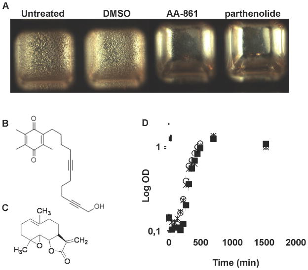
), AA-861 (□) or parthenolide ( formula image
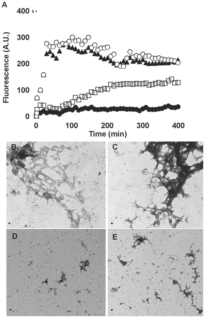
). (B–D) The molecules reduce the aggregation of New1 yeast prion expressed in E. coli cells. (B) Fluorescence and phase contrast microscopy images were taken of E. coli cells expressing the yeast prion domain New1 fused to CFP and induced with 5 μM IPTG (upper panel). A decrease in the number of cells accumulating New1-CFP foci was observed after treatment with 120 μM of (middle panel) AA-861 and (lower panel) parthenolide. (C) The number of cells accumulating New1-CFP foci was quantified and expressed as percentage of cells with foci, and (D) the average intensity of these foci expressed as arbitrary units.

Comment in

References

    1. Amijee H, Madine J, Middleton DA, Doig AJ. Inhibitors of protein aggregation and toxicity. Biochem Soc Trans. 2009;37:692–696. - PubMed
    1. Badtke MP, Hammer ND, Chapman MR. Functional amyloids signal their arrival. Sci Signal. 2009;2:pe43. - PMC - PubMed
    1. Blanco LP, Evans ML, Smith DR, Badtke MP, Chapman MR. Diversity, biogenesis and function of microbial amyloids. Trends Microbiol. 2012;20:66–73. - PMC - PubMed
    1. Branda SS, Chu F, Kearns DB, Losick R, Kolter R. A major protein component of the Bacillus subtilis biofilm matrix. Mol Microbiol. 2006;59:1229–1238. - PubMed
    1. Branda SS, Gonzalez-Pastor JE, Ben-Yehuda S, Losick R, Kolter R. Fruiting body formation by Bacillus subtilis. Proc Natl Acad Sci U S A. 2001;98:11621–11626. - PMC - PubMed

Publication types

MeSH terms