. 2013 Jan 24;20(1):123-33.
doi: 10.1016/j.chembiol.2012.11.008.

Identification of widespread adenosine nucleotide binding in Mycobacterium tuberculosis

Affiliations

Identification of widespread adenosine nucleotide binding in Mycobacterium tuberculosis

Charles Ansong et al. Chem Biol. .

Abstract

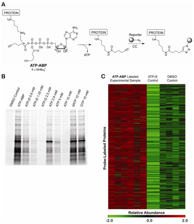
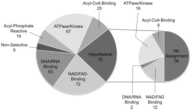
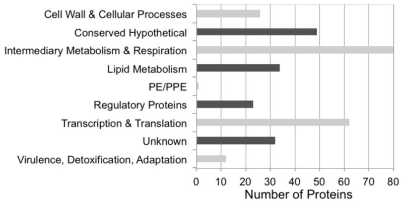
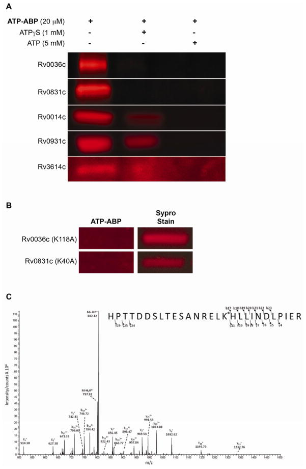
Computational prediction of protein function is frequently error-prone and incomplete. In Mycobacterium tuberculosis (Mtb), ~25% of all genes have no predicted function and are annotated as hypothetical proteins, severely limiting our understanding of Mtb pathogenicity. Here, we utilize a high-throughput quantitative activity-based protein profiling (ABPP) platform to probe, annotate, and validate ATP-binding proteins in Mtb. We experimentally validate prior in silico predictions of >240 proteins and identify 72 hypothetical proteins as ATP binders. ATP interacts with proteins with diverse and unrelated sequences, providing an expanded view of adenosine nucleotide binding in Mtb. Several hypothetical ATP binders are essential or taxonomically limited, suggesting specialized functions in mycobacterial physiology and pathogenicity.


Figures

Figure 1
Figure 1. Probe structure, selective labeling, and identification of Mtb proteins by ATP-ABP
(A) ATP-ABP structure and labeling of proteins. The ε-amino group of Lys residues reacts with one of two acyl phosphate moieties on the probe transferring the click-chemistry (CC) compatible alkyne unit to the protein and releasing ATP. Biotin (MS analysis) or Cy5.5 (fluorescent gel analysis) is appended to the probe-labeled protein via CC. (B) In-gel analysis of ATP-ABP labeled Mtb lysate. Proteomes were labeled with ATP-ABP (20 μM) alone and in the presence of ATPγs, ATP, dATP, and GTP. Labeled proteins were visualized after SDS-PAGE. (C) Heat map illustration of quantitative functional activity profile for 317 Mtb proteins, demonstrating reproducibility within probe-labeled sample replicates (ATP-ABP), no-probe control sample replicates (DMSO Control) and ATPγS-pretreated control sample replicates (ATPγS Control). The MS-measured protein abundances are listed in Supplemental Table S1. The abundance values were converted in MultiExperiment Viewer (MeV) (Saeed, et al., 2006) to normalized score (z-score) for visualization. The scale is MeV normalized score (z-score) from low (green) to high (red).
Figure 2
Figure 2. Specificity of ATP-ABP labeling in the Mtb proteome
Pie chart shows functional classification of 317 proteins confidently identified as interacting with the ATP-ABP based on chemistry of the ATP-ABP, literature mining, and in silico prediction. The insert pie chart shows the further functional classification of 72 hypothetical proteins identified. Approximately 45% of the hypothetical proteins were confirmed to be ATP-binding proteins by additional bioinformatics analyses.
Figure 3
Figure 3. Pathway distribution of 317 ATP-ABP labeled proteins from Mtb
Proteins were mapped into functional processes and pathways using TBDB.org gene mapping programs.
Figure 4
Figure 4. Validation of hypothetical protein labeling
(A) ATP-ABP (20 μM) labeling of recombinantly expressed hypothetical proteins Rv0036c and Rv0831c, T7SS protein Rv3614c, and Ser/Thr protein kinases Rv0014c and Rv0931c (MS spectra showing site of probe labeling are shown in Supplemental Figure S1). Labeling of the hypothetical proteins was competitively inhibited by ATPγS (1 mM) and ATP (5 mM), showing adenosine-dependent probe binding. (B) ATP-ABP (20 μM) labeling of the K118A mutant of Rv0036c, and the K40A mutant of Rv0831c, revealing no probe labeling following click chemistry addition of a Cy5.5 dye and SDS-PAGE separation of proteins. Sypro Ruby Red stain indicates total protein used for labeling. (C) Annotated experimental MS/MS spectra showing labeling at lysine 40 of the peptide, “R.HPTTDSLTESANRELK*HLLINDLPIER.Q,” from the probe-labeled hypothetical protein RV0831c.
Figure 5
Figure 5. Identification of novel Mtb H37Rv genes
Three peptides identified by MS-based proteomics map to the genomic region 1113888 to 1114109 on the forward strand, where no gene had been previously predicted by computational approaches. Note canonical start codon ATG upstream of peptides.

References

    1. Ansong C, Purvine SO, Adkins JN, Lipton MS, Smith RD. Proteogenomics: needs and roles to be filled by proteomics in genome annotation. Brief Funct Genomic Proteomic. 2008;7:50–62. - PubMed
    1. Barglow KT, Cravatt BF. Activity-based protein profiling for the functional annotation of enzymes. Nat Methods. 2007;4:822–827. - PubMed
    1. Bork P. Powers and pitfalls in sequence analysis: the 70% hurdle. Genome Res. 2000;10:398–400. - PubMed
    1. Bork P, Koonin EV. Predicting functions from protein sequences--where are the bottlenecks? Nat Genet. 1998;18:313–318. - PubMed
    1. Cravatt BF, Wright AT, Kozarich JW. Activity-based protein profiling: from enzyme chemistry to proteomic chemistry. Annu Rev Biochem. 2008;77:383–414. - PubMed

Publication types