. 2013 Jan 23;77(2):243-50.
doi: 10.1016/j.neuron.2012.01.034.

Lovastatin corrects excess protein synthesis and prevents epileptogenesis in a mouse model of fragile X syndrome

Affiliations

Lovastatin corrects excess protein synthesis and prevents epileptogenesis in a mouse model of fragile X syndrome

Emily K Osterweil et al. Neuron. .

Abstract

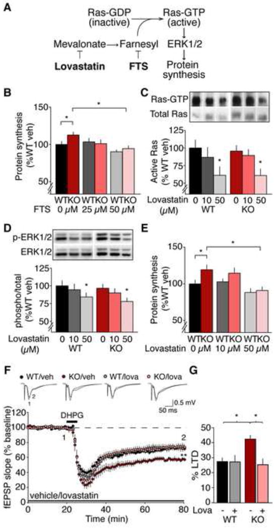
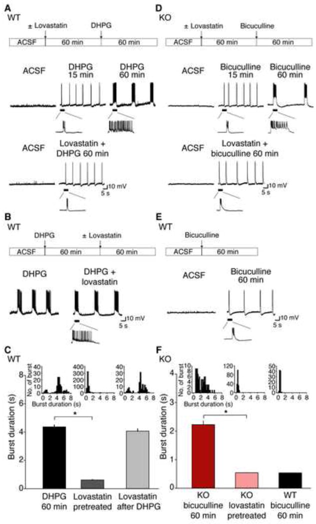
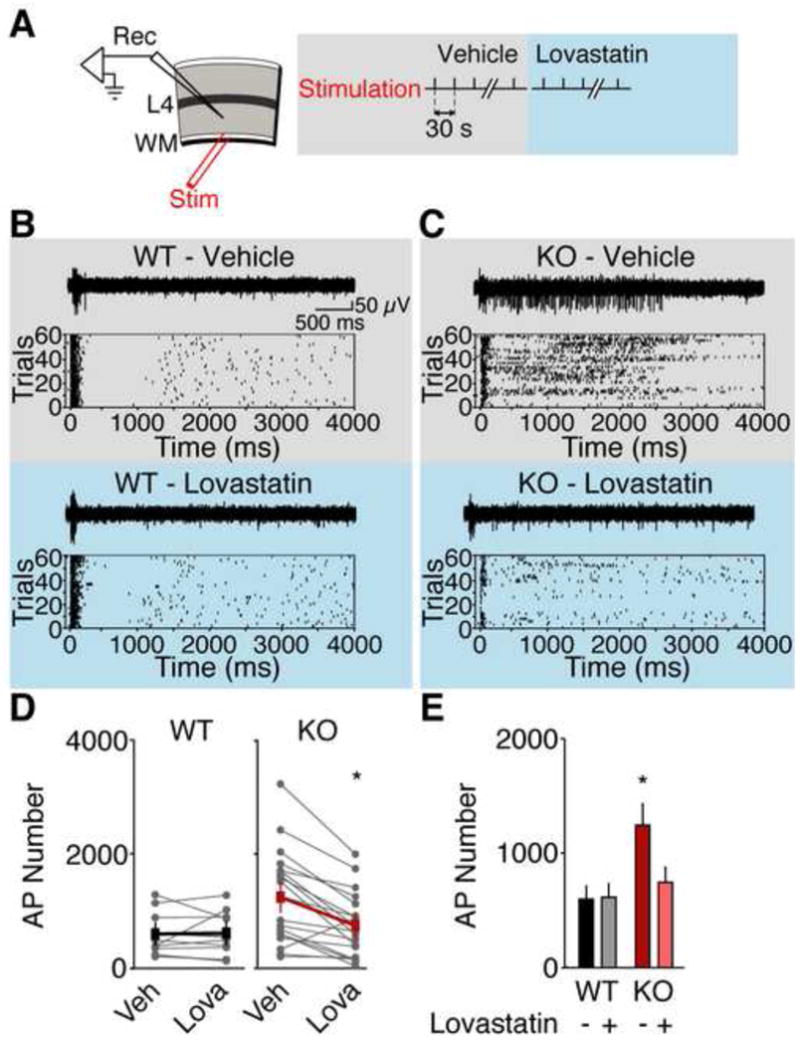
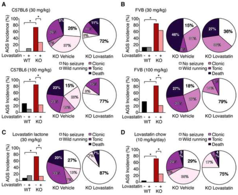
Many neuropsychiatric symptoms of fragile X syndrome (FXS) are believed to be a consequence of altered regulation of protein synthesis at synapses. We discovered that lovastatin, a drug that is widely prescribed for the treatment of high cholesterol, can correct excess hippocampal protein synthesis in the mouse model of FXS and can prevent one of the robust functional consequences of increased protein synthesis in FXS, epileptogenesis. These data suggest that lovastatin is potentially disease modifying and could be a viable prophylactic treatment for epileptogenesis in FXS.


Conflict of interest statement

Competing Interests Statement: Mark Bear discloses a financial interest in Seaside Therapeutics.

Figures

Figure 1
Figure 1
Lovastatin inhibits Ras-ERK1/2 signaling, normalizes excessive protein synthesis, and corrects exaggerated mGluR-LTD in the Fmr1 KO hippocampus. (A) Model of the mechanism by which lovastatin and FTS reduce Ras-ERK1/2 activation and normalize protein synthesis in the Fmr1 KO. (B) Application of FTS corrects excessive protein synthesis in the Fmr1 KO (WT: veh 100 ± 4%, 25 μM 104 ± 5%; 50 μM 90 ± 2%; KO: veh 113 ± 4%, 25 μM 101 ± 5%; 50 μM 94 ± 4%; ANOVA treatment *p = 0.0017; WT vs. KO veh *p = 0.041, KO veh vs. 50 μM *p = 0.00002; n = 15). A small but significant reduction of protein synthesis is also observed with 50 μM FTS in WT (*p = 0.0316). (C) Lovastatin inhibits Ras activation in hippocampal slices (WT: veh 100 ± 11%, 10 μM 87 ± 14%; 50 μM 61 ± 12%; KO: veh 96 ± 8%, 10 μM 89 ± 8%; 50 μM 61 ± 9%; ANOVA treatment *p = 0.0266; WT *p = 0.039, KO *p = 0.015; n = 6). (D) Lovastatin downregulates ERK1/2 (WT: veh 100 ± 7%, 10 μM 94 ± 8%; 50 μM 84 ± 5%; KO: veh 97 ± 5%, 10 μM 90 ± 6%; 50 μM 78 ± 5%; ANOVA treatment *p = 0.0037; WT *p = 0.035, KO *p = 0.008; n = 15). (E) Lovastatin normalizes protein synthesis in Fmr1 KO hippocampal slices (WT: veh 100 ± 5%, 10 μM 103 ± 4%; 50 μM 88 ± 5%; KO: veh 119 ± 6%, 10 μM 115 ± 7%; 50 μM 91 ± 4%; ANOVA genotype x treatment *p = 0.0332; WT vs. KO veh *p = 0.019, KO veh vs. 50 μM *p = 0.011; n = 11). (F) LTD was induced with 50 μM R,S-DHPG and extracellular recordings were performed in area CA1. In the presence of vehicle, greater LTD is observed in the Fmr1 KO versus WT (WT veh 72.5 ± 2.5%, KO veh 57.5 ± 2.5%, *p = 0.005, n = 9–10). 50 μM lovastatin significantly reduces LTD magnitude in the Fmr1 KO to WT levels (WT lova 72.7 ± 4.4%, KO lova 74.5 ± 3.4%; KO veh vs. lova *p < 0.001, n =11–13), but has no significant effect on LTD in the WT (WT veh vs. lova p = 0.869). Field potential traces are averages of all experiments, and were taken at times indicated by numerals; Scale bars = 0.5 mV, 5 ms. (G) Lovastatin significantly reduces LTD magnitude in the Fmr1 KO to WT levels (ANOVA genotype x treatment *p = 0.021). LTD magnitude was assessed by a comparison of the averaged last 5 minutes pre-DHPG and the last 5 min of recordings (minutes 55–60 post-DHPG). N = animals. Error bars = s.e.m.
Figure 2
Figure 2. Lovastatin blocks mGluR-mediated epileptiform bursting in the Fmr1 KO hippocampus
Intracellular recordings were performed on CA3 pyramidal neurons in WT and Fmr1 KO hippocampal slices. (A) In WT slices, 50 μM R,S-DHPG leads to bursting from CA3 neurons, which progresses to epileptiform discharges by 60 min. This epileptiform activity is prevented by a 60 min pre-treatment with 50 μM lovastatin. (B) Addition of lovastatin after 60 min post-DHPG did not affect the duration of epileptiform discharges. (C) Mean epileptiform burst durations from WT slices under the following conditions: at 60 min post-DHPG (4.359 ± 0.14 sec; n = 125 discharges, 6 slices from 6 animals), lovastatin-pretreated (0.618 ± 0.03 sec; n = 545 discharges, 12 slices from 7 animals), and lovastatin after DHPG (4.066 ± 0.15 sec; n = 124 discharges, 5 slices from 5 animals). Slices pretreated with lovastatin showed significant reduction in burst duration compared to vehicle-pretreated slices (*p = 0.0000218) while there was no significant difference in burst duration between DHPG 60 min and lovastatin after DHPG (p = 0.072). (D) In Fmr1 KO slices, synaptic mGluR activation by spontaneous activity in bicuculline induces prolonged epileptiform discharges, which are blocked with 60 min pre-treatment with 50 μM lovastatin. (E) In contrast to Fmr1 KO slices, bicuculline fails to induce prolonged epileptiform discharges in WT slices. (F) Plot of mean epileptiform burst durations from Fmr1 KO slices 60 min post-bicuculline ± 50 μM lovastatin, and WT slices 60 min post-bicuculline. Fmr1 KO slices pretreated with lovastatin showed significant reduction in burst duration (0.528 ± 0.01 sec; n = 182 discharges; 10 slices from 5 animals) compared to vehicle-pretreated slices (2.227 ± 0.13 sec; n = 90 discharges; 8 slices from 6 animals; *p = 0.000022) while there was no significant difference in burst duration between lovastatin pretreated Fmr1 KO slices and bicuculline treated WT slices (0.537 ± 0.01 sec; n = 88 discharges; 5 slices from 5 animals; p = 0.995). Insets indicate summary frequency histograms of synchronized discharges in each experimental condition. Error bars = s.e.m.
Figure 3
Figure 3. Lovastatin reduces excitability in the Fmr1 KO visual cortex
(A) Extracellular recordings were performed in layer 5 of visual cortical slices. Trains of action potentials (APs) were evoked with 60 trials of white matter stimulation. Responses were collected in ACSF plus vehicle, then ACSF plus 50 μM lovastatin. Representative traces and raster plots of recordings from WT (B) and Fmr1 KO (C) Prolonged firing in the Fmr1 KO is corrected with 50 μM lovastatin application. (D) Lovastatin significantly reduces firing in Fmr1 KO but not WT visual cortical slices. (E) The mean number of action potentials is significantly higher in Fmr1 KO slices versus WT slices, and 50 μM lovastatin restores normal firing to the Fmr1 KO slices (WT veh 100 ± 20%, WT lova 103 ± 20%, KO veh 203 ± 44%, KO lova 113 ±29%; ANOVA genotype x treatment *p = 0.0022; WT vs. KO veh *p = 0.0258, KO veh vs. lova *p = 0.0001, WT veh vs. lova p = 0.8412; WT n = 10 slices from 5 animals, KO n = 19 slices from 9 animals). Error bars represent s.e.m.
Figure 4
Figure 4. Lovastatin significantly reduces AGS incidence and severity in the Fmr1 KO
Fmr1 KO and WT mice on the C57BL/6 (A,C,D) or FVB (B) backgrounds were treated as indicated, tested for AGS, and scored for wild running, clonic seizure, tonic seizure, and death. (A) Injection of 30 mg/kg lovastatin acid significantly reduces AGS incidence in the Fmr1 KO on the C57BL/6 background (*p = 0.009; n = 18–19). Pie charts show a significant shift in the severity distribution of Fmr1 KO mice treated with vehicle vs. 30 mg/kg lovastatin (WT vs. KO veh *p = 0.002, WT vs. KO lov p = 0.999; KO veh vs. lov *p = 0.041). Injection of a higher100 mg/kg dose of lovastatin also significantly reduces AGS incidence (*p = 0.005; n = 13) and attenuates AGS severity (KO veh vs. lov *p = 0.015; WT vs. KO veh. *p = 0.04; WT vs. KO lov p = 0.999) in Fmr1 KO mice on the C57BL/6 background. (B) In Fmr1 KO mice bred on the FVB background, injection of 30 mg/kg lovastatin does not significantly reduce AGS incidence (KO veh 85%, KO lov 64%, p = 0.357) or severity (KO veh vs. lov p = 0.862), however a higher dose of 100 mg/kg lovastatin significantly reduces both AGS incidence (KO veh 82%, KO lov 21%, *p = 0.005; n = 11–14) and severity (KO veh vs. lov *p = 0.022; WT vs. KO veh *p = 0.016; WT vs. KO lov p = 0.999). (C) Injection of 30 mg/kg lovastatin in the lactone form significantly reduces AGS incidence (*p = 0.009; n = 15) and severity (KO veh vs. lov *p = 0.009; WT vs. KO veh *p = 0.005; WT vs. KO lov p = 0.999) in Fmr1 KO mice. (D) 48 h feeding of 0.01% lovastatin chow significantly reduces AGS incidence (KO con 71%, KO lov 25%, *p = 0.003; n = 21–24) and severity (KO veh vs. lov *p = 0.016; WT vs. KO veh *p = 0.0001; WT vs. KO lov p = 0.461) in Fmr1 KO mice. N = animals.

Comment in

References

    1. Bear MF, Huber KM, Warren ST. The mGluR theory of fragile X mental retardation. Trends Neurosci. 2004;27:370–377. - PubMed
    1. Berry-Kravis E. Epilepsy in fragile X syndrome. Dev Med Child Neurol. 2002;44:724–728. - PubMed
    1. Bhakar AL, Dolen G, Bear MF. The Pathophysiology of Fragile X (and What It Teaches Us about Synapses) Annu Rev Neurosci. 2012;35:417–443. - PMC - PubMed
    1. Bianchi R, Chuang SC, Zhao W, Young SR, Wong RK. Cellular plasticity for group I mGluR-mediated epileptogenesis. J Neurosci. 2009;29:3497–3507. - PMC - PubMed
    1. Choi CH, Schoenfeld BP, Bell AJ, Hinchey P, Kollaros M, Gertner MJ, Woo NH, Tranfaglia MR, Bear MF, Zukin RS, et al. Pharmacological reversal of synaptic plasticity deficits in the mouse model of fragile X syndrome by group II mGluR antagonist or lithium treatment. Brain Res. 2011;1380:106–119. - PMC - PubMed

Publication types