. 2013 Jun;62(6):861-72.
doi: 10.1016/j.metabol.2012.12.012. Epub 2013 Jan 24.

Renin inhibition and AT(1)R blockade improve metabolic signaling, oxidant stress and myocardial tissue remodeling

Affiliations

Renin inhibition and AT(1)R blockade improve metabolic signaling, oxidant stress and myocardial tissue remodeling

Adam Whaley-Connell et al. Metabolism. 2013 Jun.

Abstract

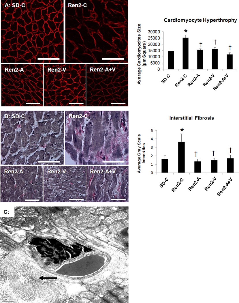
Objective: Strategies that block angiotensin II actions on its angiotensin type 1 receptor or inhibit actions of aldosterone have been shown to reduce myocardial hypertrophy and interstitial fibrosis in states of insulin resistance. Thereby, we sought to determine if combination of direct renin inhibition with angiotensin type 1 receptor blockade in vivo, through greater reductions in systolic blood pressure (SBP) and aldosterone would attenuate left ventricular hypertrophy and interstitial fibrosis to a greater extent than either intervention alone.

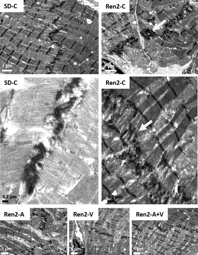
Materials/methods: We utilized the transgenic Ren2 rat which manifests increased tissue expression of murine renin which, in turn, results in increased renin-angiotensin system activity, aldosterone secretion and insulin resistance. Ren2 rats were treated with aliskiren, valsartan, the combination (aliskiren+valsartan), or vehicle for 21 days.

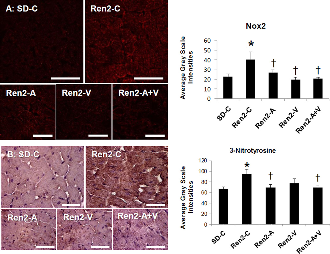
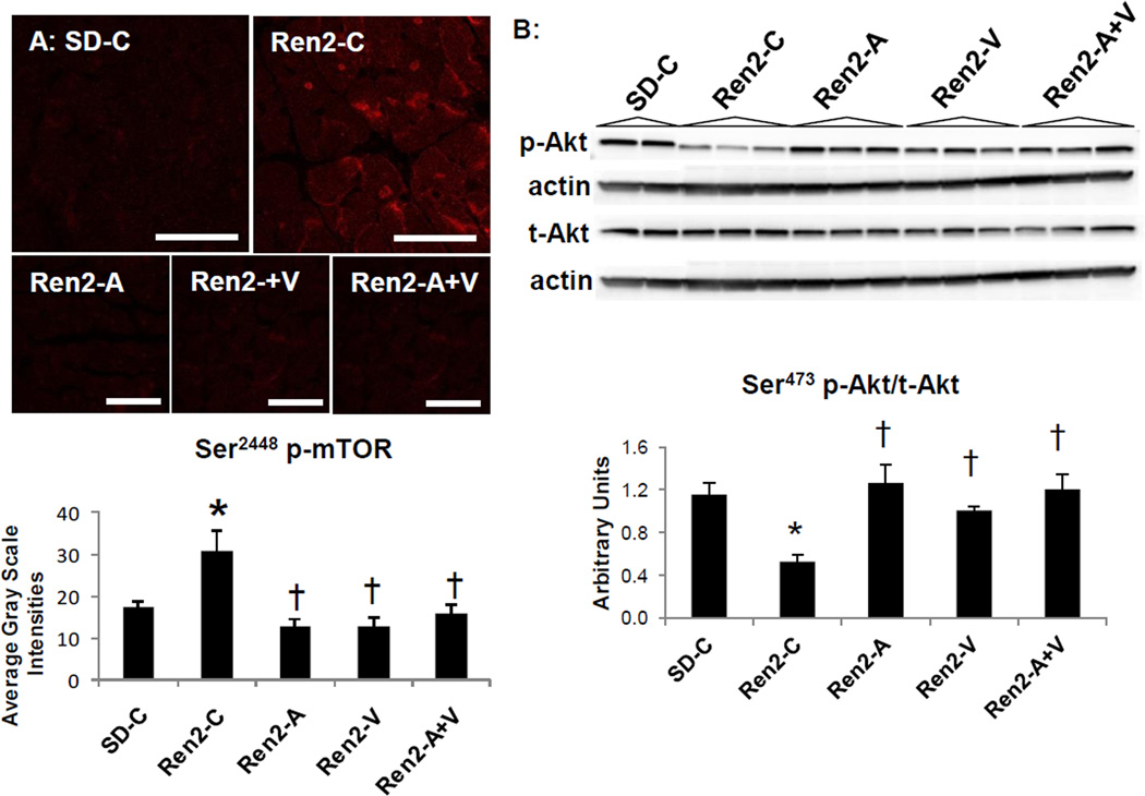
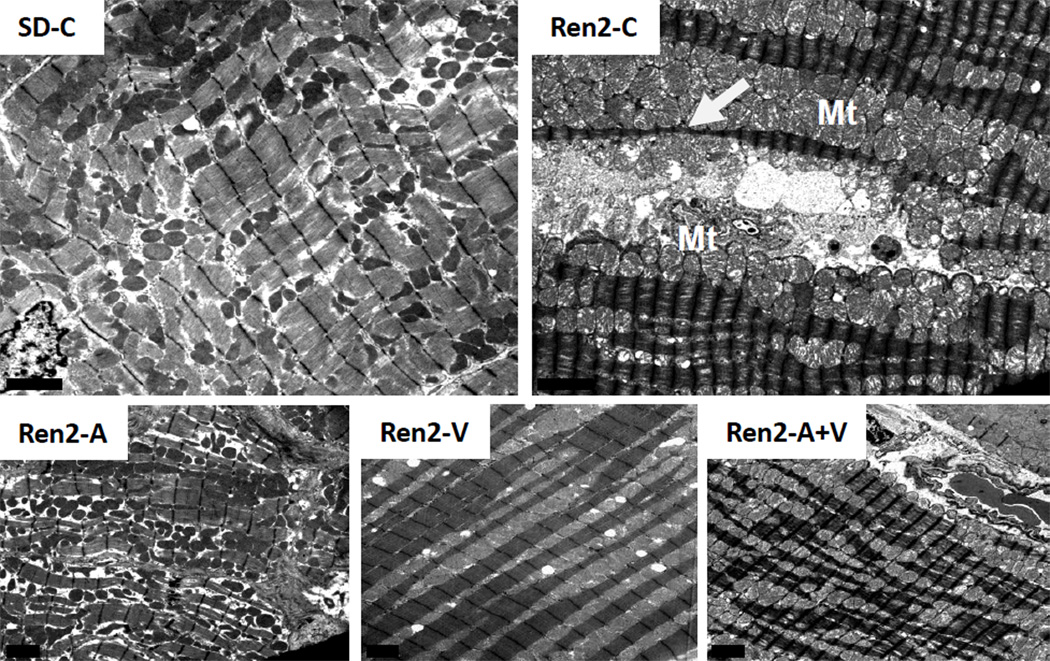
Results: Compared to Sprague-Dawley controls, Ren2 rats displayed increased systolic blood pressure, elevated serum aldosterone levels, cardiac tissue hypertrophy, interstitial fibrosis and ultrastructural remodeling. These biochemical and functional alterations were accompanied by increases in the NADPH oxidase subunit Nox2 and 3-nitrotyrosine content along with increases in mammalian target of rapamycin and reductions in protein kinase B phosphorylation. Combination therapy contributed to greater reductions in systolic blood pressure and serum aldosterone but did not result in greater improvement in metabolic signaling or markers of oxidative stress, fibrosis or hypertrophy beyond either intervention alone.

Conclusions: Thereby, our data suggest that the greater impact of combination therapy on reductions in aldosterone does not translate into greater reductions in myocardial fibrosis or hypertrophy in this transgenic model of tissue renin overexpression.


Figures

Figure 1
Figure 1. Echocardiography measures in the Ren2 Rat
A) Representative images of m-mode echocardiography of Ren2 controls (Ren2-C) compared to age-matched Sprague-Dawley controls (SD-C) with B) LV wall dimensional and ejection fraction measures below. Values presented as mean ± standard error. *, p<0.05 when Ren2-C are compared to SD-C; †, p<0.05 when Ren2 rats treated with either aliskiren (Ren2-A), valsartan (Ren2-V), or combination (Ren2-A+V) are compared to age-matched Ren2-C.
Figure 2
Figure 2. Markers of cardiac tissue hypertrophy and fibrosis in the Ren2 Rat
A) Representative images of semi-quantitative analysis of heme-pomatia agglutinin with measured to the right. B) Verhoeff-Van Gieson (VVG) stain for elastin and collagen with measures of tubulointerstitial fibrosis to the right. Values presented as mean ± standard error. *, p<0.05 when Ren2 controls (Ren2-C) are compared to age-matched Sprague-Dawley controls (SD-C); †, p<0.05 when Ren2 rats treated with either aliskiren (Ren2-A), valsartan (Ren2-V), or combination (Ren2-A+V) are compared to age-matched Ren2-C. C) Ultrastructural analysis utilizing transmission electron microscopy of the Ren2 demonstrate organized fibrillar collagen in the interstitial and perivascular regions. This image demonstrates the marked pericapillary interstitial organized fibrillar collagen (arrow) typical of early ultrastructural fibrosis that appears asteroid-like shaped around a 4×6 µm interstitial capillary. Scale bar = 1 µm.
Figure 3
Figure 3. Markers of oxidant stress in the Ren2 Rat
A) Representative confocal images of NADPH oxidase subunit Nox2 with measures of average grey scale intensities to the right. B) Representative images of 3-nitrotrysoine with corresponding measures to the right. Values presented as mean ± standard error. *, p<0.05 when Ren2 controls (Ren2-C) are compared to age-matched Sprague-Dawley controls (SD-C); †, p<0.05 when Ren2 rats treated with either aliskiren (Ren2-A), valsartan (Ren2-V), or combination (Ren2-A+V) are compared to age-matched Ren2-C. Scale bar = 50 µm.
Figure 4
Figure 4. Kinases in cardiac growth in the Ren2 rat
A) Representative images from immunohistochemistry analysis of Serine (Ser) p-mTOR at 2248 with corresponding measures below. Scale bar = 50 µm. B) Western blot analysis of phosphorylated (p) Akt at Threonine (Thr) 389 compared to total Akt in cytosolic fractions with densitometry analysis of pAkt/tAkt below. Values presented as mean ± standard error. *, p<0.05 when Ren2 controls (Ren2-C) are compared to age-matched Sprague-Dawley controls (SD-C); †, p<0.05 when Ren2 rats treated with either aliskiren (Ren2-A), valsartan (Ren2-V), or combination (Ren2-A+V) are compared to age-matched Ren2-C.
Figure 5
Figure 5. Mitochondrial Content in the Ren2 Heart
Representative images from ultrastructural analysis of the Ren2 myocardium utilizing transmission electron microsopy. In cross section of control transgenic Ren2 rats (Ren2-C; top right panel), there are excessive numbers of mitochondria (Mt) which appear to morphologically compress the sarcoplasmic reticulum (SR) structures (white arrow). The cristae are often absent and typically the mitochondria matrix is lost and appears more electron lucent. Notably, there is also thinning and disorganization of sarcomeres. These observations are not observed in Sprague Dawley control (SD-C) rats and restored in treated Ren2 rats to similar extent with aliskiren (Ren2-A), valsartan (Ren2-V), and combination treatment with both aliskiren and valsartan (Ren2-A+V). Scale bar = 2 µm.
Figure 6
Figure 6. Myocardial tissue remodeling in the Ren2 Heart
Representative images from ultrastructural analysis of the Ren2 myocardium utilizing transmission electron microscopy. In cross section of control transgenic Ren2 rats (Ren2-C; top right panel), the intercalated discs appear to be elongated with increased convolutions in order to increase surface areas between two cardiomyocytes (white arrow). Scale bar = 1 µm. This results in the appearance of being duplicated (middle panels). Scale bar = 0.2 µm. These observations are not observed in Sprague Dawley control (SD-C) rats and restored in treated Ren2 rats to similar extent with aliskiren (Ren2-A), valsartan (Ren2-V), and combination treatment.

Similar articles

Cited by

References

    1. Sowers JR, Whaley-Connell A, Epstein M. The emerging clinical implications of the role of aldosterone in the metabolic syndrome and resistant hypertension. Ann Intern Med. 2009;150:776–783. - PMC - PubMed
    1. Botdorf J, Chaudhary K, Whaley-Connell A. Hypertension in cardiovascular and kidney disease. Cardiorenal Med. 2011;1:183–192. - PMC - PubMed
    1. Reinhard H, Hansen PR, Wiinberg N, et al. NT-proBNP, echocardiographic abnormalities and subclinical coronary artery disease in high risk type 2 diabetic patients. Cardiovasc Diabetol. 2012;11:19–19. - PMC - PubMed
    1. Parving HH, Brenner BM, McMurray JJ, et al. Aliskiren Trial in Type 2 Diabetes Using Cardio-Renal Endpoints (ALTITUDE): rationale and study design. Nephrol Dial Transplant. 2009;2009;24:1663–1671. - PubMed
    1. Pulakat L, Demarco VG, Whaley-Connell A, et al. The impact of over-nutrition on insulin metabolic signaling in the heart and the kidney. Cardiorenal Med. 2011;1(2):102–112. - PMC - PubMed

Publication types

MeSH terms