. 2013 Feb 7;92(2):293-300.
doi: 10.1016/j.ajhg.2012.12.014. Epub 2013 Jan 24.

Mutations in DNA2 link progressive myopathy to mitochondrial DNA instability

Affiliations

Mutations in DNA2 link progressive myopathy to mitochondrial DNA instability

Dario Ronchi et al. Am J Hum Genet. .

Abstract

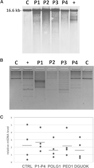
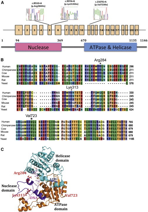
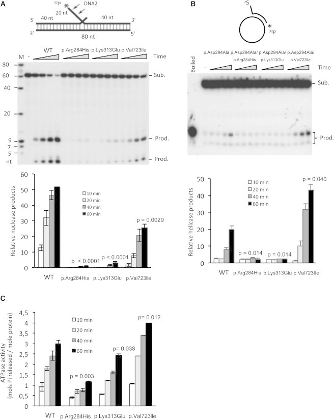
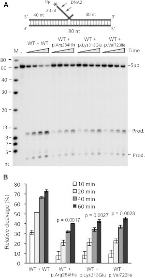
Syndromes associated with multiple mtDNA deletions are due to different molecular defects that can result in a wide spectrum of predominantly adult-onset clinical presentations, ranging from progressive external ophthalmoplegia (PEO) to multisystemic disorders of variable severity. The autosomal-dominant form of PEO is genetically heterogeneous. Recently, causative mutations have been reported in several nuclear genes that encode proteins of the mtDNA replisome machinery (POLG, POLG2, and C10orf2) or that are involved in pathways for the synthesis of deoxyribonuclotides (ANT1 and RRM2B). Despite these findings, putative mutations remain unknown in half of the subjects with PEO. We report the identification, by exome sequencing, of mutations in DNA2 in adult-onset individuals with a form of mitochondrial myopathy featuring instability of muscle mtDNA. DNA2 encodes a helicase/nuclease family member that is most likely involved in mtDNA replication, as well as in the long-patch base-excision repair (LP-BER) pathway. In vitro biochemical analysis of purified mutant proteins revealed a severe impairment of nuclease, helicase, and ATPase activities. These results implicate human DNA2 and the LP-BER pathway in the pathogenesis of adult-onset disorders of mtDNA maintenance.


Figures

Figure 1
Figure 1
Molecular Findings in Muscle Tissue of Probands with DNA2 Mutations (A) Southern-blot analysis of muscle mtDNA derived from probands 1–4 revealed the presence of multiple bands. “C” indicates control samples that display only a single band corresponding to WT mtDNA, and “+” indicates a positive control (POLG-mutated subject). (B) Long PCR. This panel shows an agarose-electrophoresis separation of the WT mtDNA long-PCR amplified fragment (approximately 8,520 bp). Variably abundant extrabands due to mtDNA-deleted molecules from muscle DNA are observed in all the probands, but not in age-matched controls (“C”). (C) Quantitative PCR analysis of mtDNA content in patients’ muscle. Real-time quantitative PCR analysis was performed with primers and probes for human MTCYB (MIM 516020, mtDNA) and APP (MIM 104760, nDNA). Primer sequences and PCR conditions are available upon request. Results were analyzed with a Student’s t test. The mtDNA copy number was determined by quantitative real-time PCR in skeletal-muscle samples of our probands (P1–P4, n = 4), age-matched control individuals (n = 4), and three groups of individuals with autosomal-dominant or -recessive PEO and mutations in POLG (n = 4), PEO1 (n = 4), or DGUOK (n = 4); no significant difference was observed. All determinations were performed in triplicate. Values are normalized to a control sample. Horizontal bars indicate mean values.
Figure 2
Figure 2
DNA2 Variants Occur at Conserved Residues Located in Important Structural Elements of Human DNA2 (A) A scheme of DNA2 includes the location of the identified mutations in the coding sequence. Exons are numbered. A Diagram representing human DNA2 shows the functional domains conserved in this enzyme. (B) Multiple-sequence alignment of nuclease and ATPase domains for the DNA2 mammalian and yeast orthologous protein sequences with the use of CLC Bio Main Workbench v.5.1. The tabular format of six orthologs of DNA2 sequences is color coded to display the conservation at each position in the alignment. (C) Homology model of DNA2 with ADP (blue) binding in the catalytic center of the ATPase domain (brown) interacts with ssDNA (pink). The positions of three residues, Arg284, Lys313, and Val723, are illustrated as red sticks. The nuclease and helicase domains are displayed in purple and cyan, respectively.
Figure 3
Figure 3
Changes in Enzymatic Activities of Altered Forms of DNA2 (A) Endonuclease activities of WT and altered forms of DNA2. In the top panel is the DNA double-flap substrate. WT, p.Arg284His, p.Lys313Glu, or p.Val723Ile human DNA2 (0.8 nM) was incubated with 0.25 pmol 5′ end 32P-labeled flap DNA substrate. Reactions were carried out at 37°C for 10, 20, 40, and 60 min. Arrows indicate the cleavage sites. In the bottom panel is the quantification of DNA2 endonuclease activity. Error bars indicate SEM. (B) Helicase activities of WT and altered forms of DNA2. In the top panel is the DNA helicase substrate. DNA2 was first altered for the elimination of nuclease activity (p.Asp294Ala) so that helicase activity could be assayed. A helicase mixture containing 1 fmol 3′-end-labeled duplex substrate and 100 nM purified recombinant p.Asp294Ala, p.Asp294Ala/p.Arg284His, p.Asp294Ala/p.Lys313Glu, or p.Asp294Ala/p.Val723Ile DNA2 was incubated at 37°C for 10, 20, 40, and 60 min. In the bottom panel is the quantification of DNA2 helicase activity. Error bars indicate SEM. (C) ATPase activity of WT and altered forms of DNA2. ATPase activity of WT, p.Arg284His, p.Lys313Glu, or p.Val723Ile forms of human DNA2 was measured with an ATPase assay kit. A reaction mixture containing 30 nM recombinant protein and 1 μg ssDNA was incubated at 37°C for 10, 20, 40, and 60 min. In each panel, the p value was calculated by a Student’s t test. Error bars indicate SEM.
Figure 4
Figure 4
Heterogeneity Effects of the Nuclease Activity of Altered Forms of DNA2 (A) 0.2 nM WT DNA2 was mixed with 0.2 nM WT or p.Arg284His, p.Lys313Glu, or p.Val723Ile forms of DNA2. The DNA2 mixture was incubated with 0.25 pmol 5′ end 32P-labeled flap DNA substrates. Reactions were carried out at 37°C for 10, 20, 40, and 60 min. (B) Quantification of DNA2 cleavage products. Values are means ± SEM of three assays. In each panel, the p value was calculated by a Student’s t test.

References

    1. Wanrooij S., Falkenberg M. The human mitochondrial replication fork in health and disease. Biochim. Biophys. Acta. 2010;1797:1378–1388. - PubMed
    1. Stumpf J.D., Copeland W.C. Mitochondrial DNA replication and disease: Insights from DNA polymerase γ mutations. Cell. Mol. Life Sci. 2011;68:219–233. - PMC - PubMed
    1. Sen D., Nandakumar D., Tang G.Q., Patel S.S. Human mitochondrial DNA helicase TWINKLE is both an unwinding and annealing helicase. J. Biol. Chem. 2012;287:14545–14556. - PMC - PubMed
    1. Copeland W.C. Defects in mitochondrial DNA replication and human disease. Crit. Rev. Biochem. Mol. Biol. 2012;47:64–74. - PMC - PubMed
    1. Virgilio R., Ronchi D., Hadjigeorgiou G.M., Bordoni A., Saladino F., Moggio M., Adobbati L., Kafetsouli D., Tsironi E., Previtali S. Novel Twinkle (PEO1) gene mutations in mendelian progressive external ophthalmoplegia. J. Neurol. 2008;255:1384–1391. - PubMed

Publication types