Review
. 2013 Apr;38(4):177-83.
doi: 10.1016/j.tibs.2012.12.005. Epub 2013 Jan 23.

A common mode of recognition for methylated CpG

Affiliations
Review

A common mode of recognition for methylated CpG

Yiwei Liu et al. Trends Biochem Sci. 2013 Apr.

Abstract

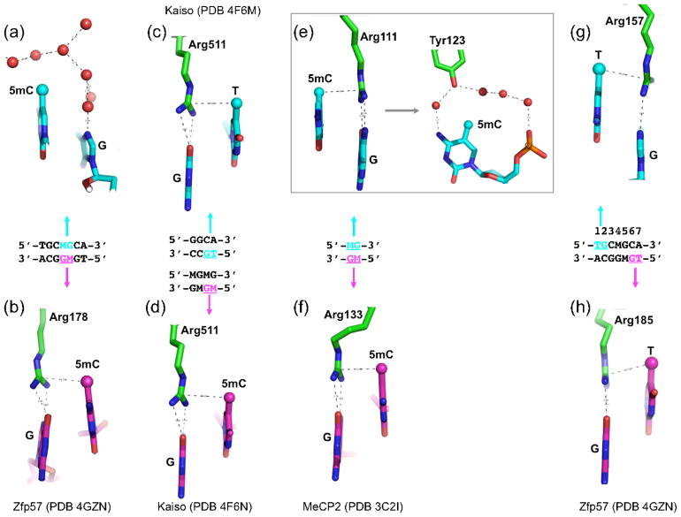
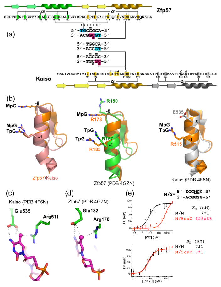
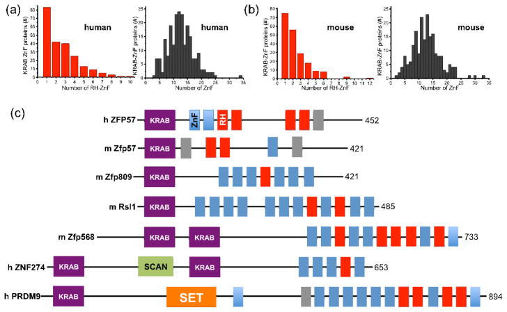
Much is known about vertebrate DNA methylation, however it is not known how methylated CpG within particular sequences is recognized. Two recent structures of C2H2 zinc finger (ZnF) proteins in complex with methylated DNA reveal a common recognition mode for 5-methylcytosine (5mC) that involves a 5mC-Arg-G triad. In the two ZnF proteins, an arginine that precedes the first Zn-binding histidine (RH motif) can interact with a 5mCpG or TpG dinucleotide. Among a family of >300 human Krüppel-associated box (KRAB) domain containing ZnF proteins examined, two-thirds contained at least one ZnF that included an RH motif. We propose that the RH-ZnF motifs provide specificity for 5mCpG, whereas the neighboring Zn fingers recognize the surrounding DNA sequence context.


Figures

Figure 1
Figure 1. A methyl-Arg-G triad forms during recognition of methyl-CpG and TpG dinucleotides in double stranded DNA
(a–b) Zfp57 recognizes the 5mCs (M) of the two DNA strands via very different interactions. The upper strand 5mC methyl is surrounded by an ordered network of water molecules (red spheres), while the lower strand 5mC methyl is part of a 5mC-Arg-G triad. (c–d) Kaiso recognizes either a specific nonmethylated DNA element containing a TpG dinucleotide (panel c) or two consecutive methylated CpG dinucleotides (panel d). In both cases, a methyl-Arg-G triad is involved. (e–f) MeCP2 forms two 5mC-Arg-G triads to symmetrically bind the palindromic fully-methylated CpG duplex. In addition, the upper strand 5mC methyl group is surrounded by an ordered network of water molecules that also includes a tyrosine hydroxyl (panel e, right). (g–h) Zfp57 uses an identical pair of thymine-Arg-G triads for the TpG dinucleotides on the two strands.
Figure 2
Figure 2. Structural comparison of Zfp57- and Kaiso-DNA interactions
(a) Structure-based sequence alignment of Zfp57 and Kaiso. The secondary structure elements (arrows for β strands and ribbons for α helices) are shown above (Zfp57) or below (Kaiso) the protein sequence. The third ZnF of Kaiso is not shown. The amino acid positions highlighted are responsible for Zn ligand binding (C2H2) and DNA base-specific interactions (−1 to −8). The arginines responsible for TpG or MpG recognition are located in the -−1 or −8 positions (relative to the first Zn-binding His). (b) (Left panel) Superimposition of the recognition helices of Zfp57 (second ZnF) and Kaiso (first ZnF). (Middle panel) Superimposition of the two helices of Zfp57. Arg150 of mouse Zfp57, at the −8 position of the first ZnF, makes a DNA phosphate interaction but is not conserved in human ZFP57. (Right panel) Superimposition of the first two helices of Kaiso. (c) In Kaiso, in addition to Arg511, the side chain of Glu535 forms a van der Waals contact with the methyl group of 5mC and one of its carboxylate oxygen atoms interacts with the N4 atom of the same 5mC base. The hydrogen bond could also be formed with the O4 atom of the thymine of TpG (presumably through protonation of the carboxylate side chain) [20]. (d) Although not aligned at the primary sequence level, Zfp57 has a spatially-conserved glutamate (Glu182) with respect to Glu535 of Kaiso. (e) DNA binding activity of Zfp57 Glu182 mutants. The 5-carboxylation (5caC) has a large effect on DNA binding by WT Zfp57, but no such effect of 5caC is seen with the E182Q (glutamate to glutamine) mutant.
Figure 3
Figure 3. KRAB-ZnF proteins containing RH-ZnF motifs
(a–b) The SysZNF database (http://lifecenter.sgst.cn/SysZNF/) was examined for KRAB (Krüppel-associated box)-ZnF proteins in human (a) or mouse (b). In each case, the distribution of KRAB-ZnF proteins containing a given number of RH-ZnF motifs is shown in the left panel and the distribution of KRAB-ZnF proteins containing a given number of ZnF repeats is shown in right panel. (c) Examples of mammalian KRAB-ZnF proteins with known biological roles. The size of each protein (in amino acids) is shown at right. The classic C2H2 ZnF motifs are shown in blue boxes and the RH-ZnF motifs are shown in red boxes. The grey boxes indicate degenerate ZnFs that include mutations affecting zinc coordination. The SCAN box, a leucine-rich region, was named after SRE-ZBP, CTfin51, AW-1 (ZNF174), and Number 18 cDNA (ZnF20) [53]. The SET domain was named after Su(var)3–9, En(zeste), and Trithorax [54].

Comment in

References

    1. Bestor T, et al. Cloning and sequencing of a cDNA encoding DNA methyltransferase of mouse cells. The carboxyl-terminal domain of the mammalian enzymes is related to bacterial restriction methyltransferases. J Mol Biol. 1988;203:971–983. - PubMed
    1. Okano M, et al. Cloning and characterization of a family of novel mammalian DNA (cytosine-5) methyltransferases. Nat Genet. 1998;19:219–220. - PubMed
    1. Tahiliani M, et al. Conversion of 5-methylcytosine to 5-hydroxymethylcytosine in mammalian DNA by MLL partner TET1. Science. 2009;324:930–935. - PMC - PubMed
    1. Ito S, et al. Tet proteins can convert 5-methylcytosine to 5-formylcytosine and 5-carboxylcytosine. Science. 2011;333:1300–1303. - PMC - PubMed
    1. He YF, et al. Tet-mediated formation of 5-carboxylcytosine and its excision by TDG in mammalian DNA. Science. 2011;333:1303–1307. - PMC - PubMed

Publication types

LinkOut - more resources