. 2013 Jan 31;152(3):570-83.
doi: 10.1016/j.cell.2013.01.003. Epub 2013 Jan 24.

Braveheart, a long noncoding RNA required for cardiovascular lineage commitment

Affiliations

Braveheart, a long noncoding RNA required for cardiovascular lineage commitment

Carla A Klattenhoff et al. Cell. .

Abstract

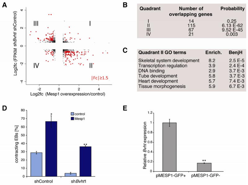
Long noncoding RNAs (lncRNAs) are often expressed in a development-specific manner, yet little is known about their roles in lineage commitment. Here, we identified Braveheart (Bvht), a heart-associated lncRNA in mouse. Using multiple embryonic stem cell (ESC) differentiation strategies, we show that Bvht is required for progression of nascent mesoderm toward a cardiac fate. We find that Bvht is necessary for activation of a core cardiovascular gene network and functions upstream of mesoderm posterior 1 (MesP1), a master regulator of a common multipotent cardiovascular progenitor. We also show that Bvht interacts with SUZ12, a component of polycomb-repressive complex 2 (PRC2), during cardiomyocyte differentiation, suggesting that Bvht mediates epigenetic regulation of cardiac commitment. Finally, we demonstrate a role for Bvht in maintaining cardiac fate in neonatal cardiomyocytes. Together, our work provides evidence for a long noncoding RNA with critical roles in the establishment of the cardiovascular lineage during mammalian development.


Figures

Figure 1
Figure 1. Identification AK143260 as a candidate heart-associated lncRNA
A. Differential expression enrichment score calculated from RNA-Seq data from biological replicates of two independent mESC lines and adult tissues (brain, liver, and skeletal muscle) representing the three germ layers. Stars represent the position of 3 example candidates and their respective Genbank accession numbers. The y-axis represents the number of lncRNAs with a particular differential expression enrichment, as denoted by each bar. B. Transcriptome analysis in a broader range of tissues showed that AK143260 was predominantly expressed in ESCs and in heart. C. Depiction of transcript structure as defined by 5′ and 3′ RACE (lower panel) was consistent with RNA sequencing reads in ESCs (upper panel). D. Northern analysis using a full-length probe for AK143260 confirms the predicted size of the transcript (~590 nucleotides) and shows reduced levels upon shRNA-mediated depletion in mESCs. E. RNA sequencing reads from rat adult heart and human adult heart samples showed no evidence of a transcript expressed in a location syntenic to AK143260. Arrows mark direction of transcription. Related to Figure S1 and Table S1.
Figure 2
Figure 2. Bvht is required for proper ESC differentiation
A. Targeting of AK143260 (Bvht) in mESCs by two independent hairpins directed to either exon 2 or 3 led to significant depletion of the transcript as measured by qRT-PCR. Experiments were performed in triplicate and error bars represent standard deviation within one representative experiment. B. Bvht depletion does not affect colony morphology and OCT4 staining in ESCs. Scale bar=100 μm. C. Percentage of spontaneously contracting EBs determined at Day 8 and 11 of differentiation (n>100 per time point). Bvht-depleted EBs showed a significant decrease in contracting EBs compared to controls. The number of resulting EBs was similar in each experiment. Experiments were performed in triplicate and error bars represent standard deviation. D. Expression of cardiac Troponin T (cTnT) measured by qRT-PCR at representative time points shows decreased levels in Bvht-depleted EBs. Error bars represent standard deviation of triplicate set of experiments. E. Antibody staining of paraffin embedded sections of Day 12 EBs showed decreased cTNT abundance in Bvht-depleted compared to controls. Scale bar=300 μm. F. Bvht-depleted EBs can form tissues representative of all three germ layers including gut-like epithelium (endoderm), cartilage (mesoderm), and neuroepithelial rosettes (ectoderm) as represented in H&E stained EB sections. Scale bar=100 μm. ** p<0.01 by two-tailed Student’s t-test. Related to Figure S2 and Table S2.
Figure 3
Figure 3. Bvht regulates a core network of genes that drive cardiac differentiation
A. Heat map representing hierarchical clustering of all genes that displayed a 3-fold or greater difference in transcript levels in Bvht-depleted EBs compared to controls at Day 0, 3, 6, and 9. B. Significant gene ontology (GO) terms retrieved by clusters G–I. C. Pearson correlation network of the EB time course generated using the Spring embedded algorithm in Cytoscape. Genes in enriched GO categories are represented in the network. A sequence of cardiac transcription factors and genes involved in myofibril organization were not induced during EB differentiation in Bvht-depleted cells. Nodes represent genes and connections represent correlation coefficient. Related to Figure S3 and S4 and Table S3.
Figure 4
Figure 4. Bvht functions upstream of MesP1
A. Gene expression comparison between Bvht depletion and Mesp1 induction. B. The overlap is highly significant for quadrants II and III as measured by hypergeometric test with Benjamini correction. Genes in quadrant II failed to activate in Bvht-depleted cells and are expressed at higher levels upon Mesp1 induction, whereas quadrant III genes displayed the opposite expression pattern. C. Enriched GO terms based on genes in quadrant II. D. Forced MesP1 expression can rescue the contraction defect in Bvht-depleted EBs. Contracting EBs were counted at Day 11 (n>100). Error bars represent standard deviation of a triplicate set of experiments. E. Bvht is enriched in cells expressing a GFP reporter under the control of the Mesp1 promoter (pMESP1-GFP) compared to GFP negative cells as analyzed by FACS at Day 5 (peak of MesP1 expression) of EB differentiation followed by qRT-PCR. Experiments were performed in triplicate and error bars represent standard deviation in all panels. ** p<0.01; * p<0.05 by two-tailed Student’s t-test.
Figure 5
Figure 5. Bvht is necessary for the transition from nascent to cardiac mesoderm in vitro
A. ESCs were differentiated into beating cardiomyocytes (CM) and progress through precardiac mesoderm (MES) and cardiac progenitor (CP) stages by sequential addition of cytokines. B. Bvht-depleted EBs were smaller than controls at Day 4 and failed to attach and organize into a monolayer at Day 5.3. Scale bar=65 μm. C. Bvht-depleted cells showed increased levels of apoptosis at Day 4 as indicated by quantification of Annexin V-positive cells, whereas no significant difference was observed at Day 2. D. Brachyury and Eomes levels remained high at Day 5.3 compared to control as measured by qRT-PCR. E., F. Core cardiac transcription factors displayed lower expression at Day 4 (E) and Day 5.3 (F) in Bvht-depleted cells compared to control. G. Expression of cell surface markers, PdgfRa and Flk-1, was significantly decreased upon Bvht depletion at Day 4 and Day 5.3 compared to control. H. EMT genes displayed altered expression patterns in Bvht-depleted-cells at Day 5.3 as measured by qRT-PCR. Values for shControl samples were normalized to 1 for each individual time point. I. Equal numbers of mCherry-shControl and GFP-shBvht1 cells were mixed and subjected to cardiac differentiation. A dramatic decrease in Bvht-depleted cells was observed at Day 4 and Day 5.3, whereas cells cultured in ESC conditions maintain equal distribution. Error bars represent standard deviation of three replicates within one representative experiment. ** p<0.01; * p<0.05 by two-tailed Student’s t-test. Related to Figure S6.
Figure 6
Figure 6. Bvht directly interacts with SUZ12 during CM differentiation
A. Biotin-RNA pull downs using full length Bvht transcript in nuclear ESC extracts showed specific binding to SUZ12. B. Deletion analysis of Bvht transcript showed that 5′ and 3′ regions were required for interaction with SUZ12. C. Bvht interacts with SUZ12 in Day 5.3 cells (CP stage) as determined by RIP using SUZ12 antibody. D. ChIP using SUZ12 antibody showed higher levels at promoter regions of cardiac and EMT genes in Bvht-depleted Day 5.3 cells compared to controls, whereas no significant changes were observed in genes expressed in other lineages (e.g. Sox1 and Sox17). Experiments were performed in triplicate and error bars represent standard deviation in all panels. ** p<0.01; * p<0.05 by two-tailed Student’s t-test. Related to Figure S7.
Figure 7
Figure 7. Bvht is required to maintain cardiac fate in neonatal cardiomyocytes
A. Bvht depleted or control nCMs were identified by the presence of a GFP reporter in the shRNA vector. Myofibrils clustered together in GFP positive control cells whereas Bvht depletion resulted in disorganized cardiac myofibril bundles. Myofibrils were visualized by IF using an antibody against cTnT (red), and GFP (green). Nuclei (blue) were stained with Hoechst. Scale bars=20 μm. B. Bvht-depleted nCMs were overall smaller than controls as assessed by the surface area of GFP positive versus control cells (n>100). C. Global levels of Bvht (qRT-PCR) in control and Bvht-depleted nCM cultures. D. Reduced expression of sarcomeric and myofibril components in Bvht-depleted nCMs as measured by qRT-PCR. E. Expression (qRT-PCR) of transcription factors involved in early cardiogenesis was increased in Bvht-depleted cells. F. Factors involved in EMT were expressed at lower levels in Bvht-depleted cells. ** p<0.01, * p<0.05 by ANOVA and Dunnett’s test.

Comment in

Similar articles

Cited by

References

    1. Banfai B, Jia H, Khatun J, Wood E, Risk B, Gundling WE, Jr, Kundaje A, Gunawardena HP, Yu Y, Xie L, et al. Long noncoding RNAs are rarely translated in two human cell lines. Genome Res. 2012;22:1646–1657. - PMC - PubMed
    1. Bernstein BE, Mikkelsen TS, Xie X, Kamal M, Huebert DJ, Cuff J, Fry B, Meissner A, Wernig M, Plath K, Jaenisch R, et al. A bivalent chromatin structure marks key developmental genes in embryonic stem cells. Cell. 2006;125:315–326. - PubMed
    1. Bondue A, Lapouge G, Paulissen C, Semeraro C, Iacovino M, Kyba M, Blanpain C. Mesp1 acts as a master regulator of multipotent cardiovascular progenitor specification. Cell Stem Cell. 2008;3:69–84. - PubMed
    1. Bondue A, Tannler S, Chiapparo G, Chabab S, Ramialison M, Paulissen C, Beck B, Harvey R, Blanpain C. Defining the earliest step of cardiovascular progenitor specification during embryonic stem cell differentiation. J Cell Biol. 2011;192:751–765. - PMC - PubMed
    1. Boyer LA, Plath K, Zeitlinger J, Brambrink T, Medeiros LA, Lee TI, Levine SS, Wernig M, Tajonar A, Ray MK, et al. Polycomb complexes repress developmental regulators in murine embryonic stem cells. Nature. 2006;441:349–353. - PubMed

Publication types

Substances

Associated data