. 2013 Apr 1;376(1):62-73.
doi: 10.1016/j.ydbio.2013.01.012. Epub 2013 Jan 23.

BMP and TGF-β pathway mediators are critical upstream regulators of Wnt signaling during midbrain dopamine differentiation in human pluripotent stem cells

Affiliations

BMP and TGF-β pathway mediators are critical upstream regulators of Wnt signaling during midbrain dopamine differentiation in human pluripotent stem cells

Jingli Cai et al. Dev Biol. .

Abstract

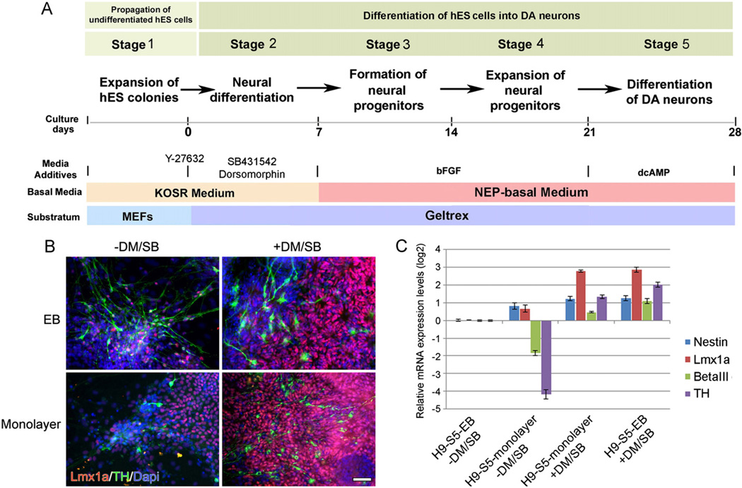
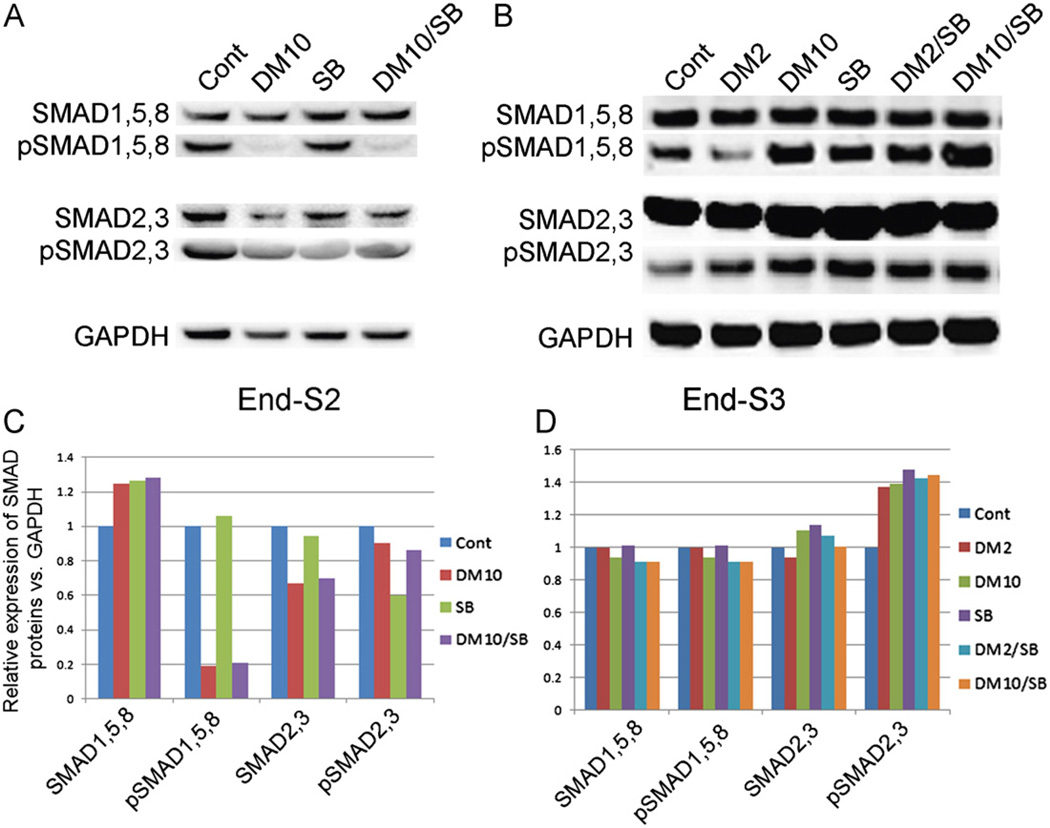
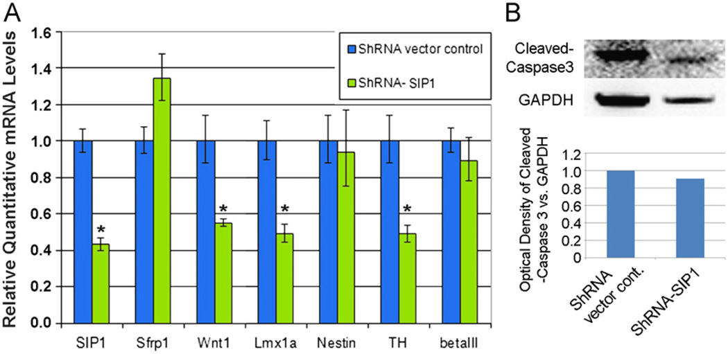
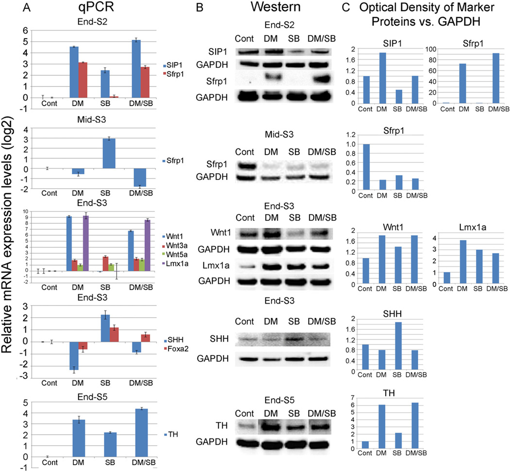
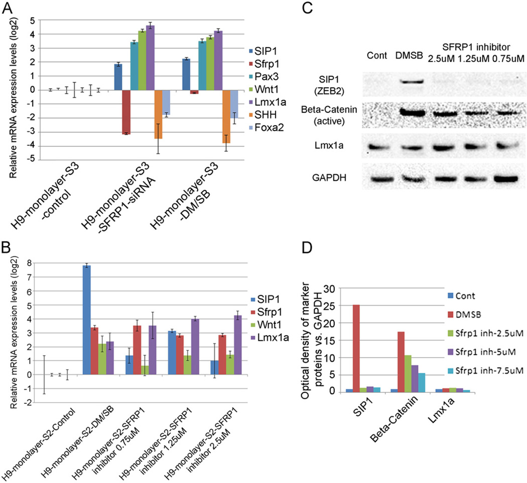
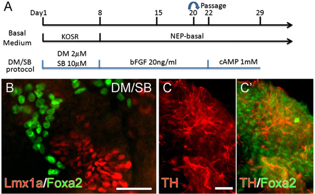
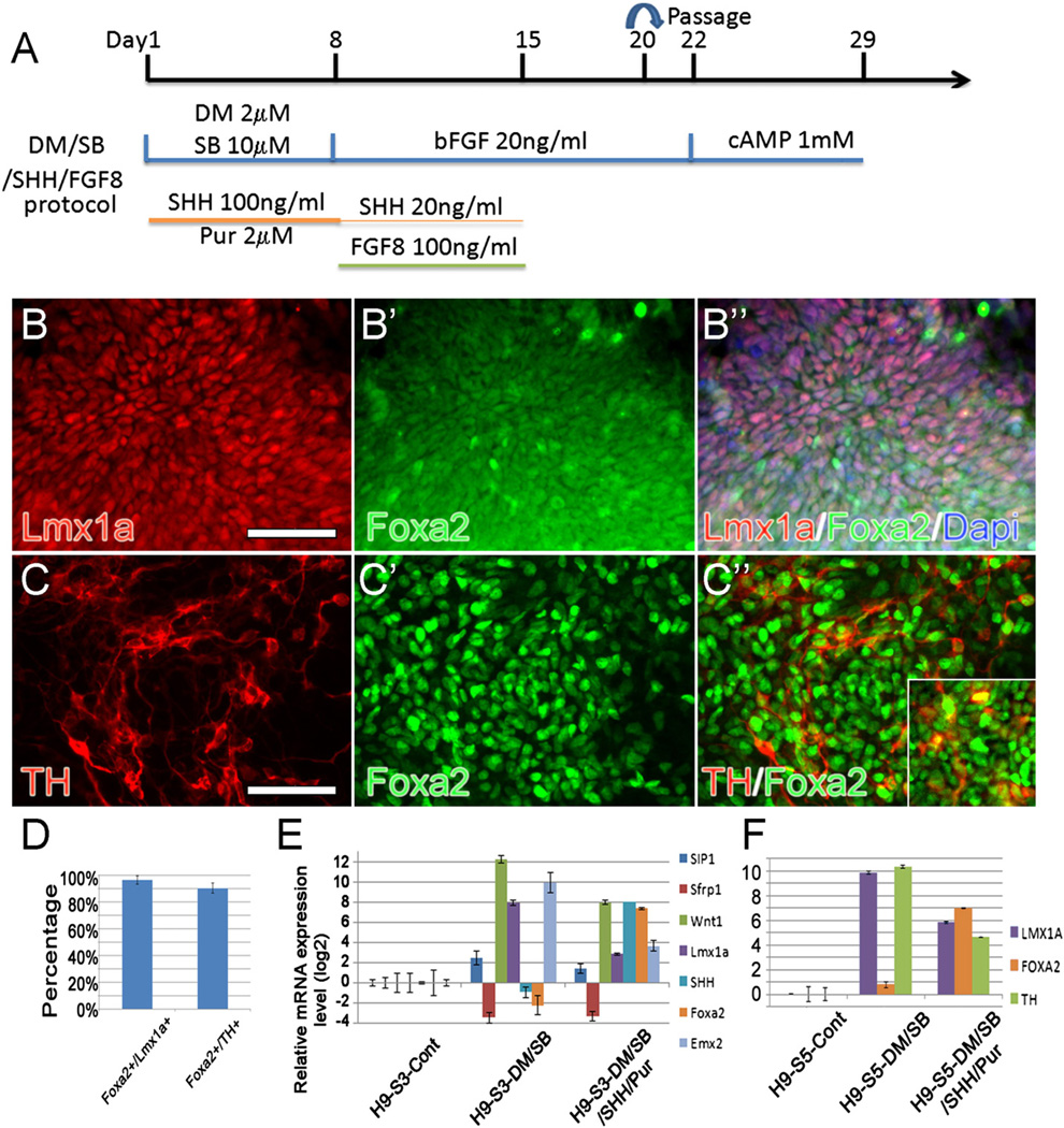
Although many laboratories currently use small molecule inhibitors of the BMP (Dorsomorphin/DM) and TGF-β (SB431542/SB) signaling pathways in protocols to generate midbrain dopamine (mDA) neurons from hES and hiPS cells, until now, these substances have not been thought to play a role in the mDA differentiation process. We report here that the transient inhibition of constitutive BMP (pSMADs 1, 5, 8) signaling, either alone or in combination with TGF-β inhibition (pSMADs 2, 3), is critically important in the upstream regulation of Wnt1-Lmx1a signaling in mDA progenitors. We postulate that the mechanism via which DM or DM/SB mediates these effects involves the up-regulation in SMAD-interacting protein 1 (SIP1), which results in greater repression of the Wnt antagonist, secreted frizzled related protein 1 (Sfrp1) in stem cells. Accordingly, knockdown of SIP1 reverses the inductive effects of DM/SB on mDA differentiation while Sfrp1 knockdown/inhibition mimics DM/SB. The rise in Wnt1-Lmx1a levels in SMAD-inhibited cultures is, however, accompanied by a reciprocal down-regulation in SHH-Foxa2 levels leading to the generation of few TH+ neurons that co-express Foxa2. If however, exogenous SHH/FGF8 is added along with SMAD inhibitors, equilibrium in these two important pathways is achieved such that authentic (Lmx1a+Foxa2+TH+) mDA neuron differentiation is promoted while alternate cell fates are suppressed in stem cell cultures. These data indicate that activators/inhibitors of BMP and TGF-β signaling play a critical upstream regulatory role in the mDA differentiation process in human pluripotent stem cells.


Figures

Fig. 1
Fig. 1
(A) Depiction of the various stages and treatments used to directly differentiate H9 cells into DA neurons in monolayer culture (modified from Iacovitti et al., 2007). (B, C) Expression levels of Lmx1a and TH are greatly increased by the treatment of stem cells with DM/SB. At stage 2, cultures were incubated with or without DM/SB then carried through the subsequent stages, (B) Stage 5 monolayer or EB-derived cultures stained for Lmx1a, TH and counter-stained with Dapi. Scale bar=50 µm, and (C) mRNA levels were quantified by qPCR compared to cells grown using the EB method in the absence of DM/SB treatment.
Fig. 2
Fig. 2
Western blot detection of protein levels of SMADs and pSMADs expressed at the end of Stg2 or Stg3 in cells treated with DM, SB or DM/SB (A, B). All cell cultures exhibit similar levels of SMADs 1, 5, 8 and SMADs 2, 3. DM and DM/SB dramatically down-regulated pSMADs 1, 5, 8 while SB only partially lowered the levels of pSMADs 2, 3. (C, D) Quantification of western blot results shown in panel A, B.
Fig. 3
Fig. 3
(A) Gene expression analysis of markers of differentiation after SIP1 knockdown at Stg1. Wnt1, Lmx1a, and TH were down-regulated at the end of stage 4 while nestin and β-III tub show no significant changes (unpaired t test): *P < 0.05. (B) Western blot detection of similar cleaved Caspase3 expression in SIP1 knockdown samples as vector control.
Fig. 4
Fig. 4
mRNA levels (A) and protein levels (B) of mDA markers examined at different stages after treatment of hES (H9 line) cells with DM, SB or DM/SB. At the end of Stg2, both SIP1 and Sfrp1 expression levels were increased after DM and DM/SB treatment. By mid-Stg3, Sfrp1 expression levels fell dramatically with DM and DM/SB treatment. At the end of Stg3, DM and DM/SB treatment greatly increased the expression of Wnt1 and Lmx1a (and somewhat increased Wnt3a and Wnt5a) while SHH expression decreased. At the end of Stg5, TH expression levels were increased with DM, SB and DM/SB treatment. (C) Quantification of Western blot results shown in panel B.
Fig. 5
Fig. 5
(A) Gene expression analysis of markers of differentiation 2 days after knockdown of Sfrp1 on Day 2 of Stg3. Sfrp1 knockdown produced similar changes in its downstream genes, Pax3, Wnt1, Lmx1a, SHH and Foxa2 as DM/SB treatment at Stg2. (B) Treatment with EMD Millipore 344300, a chemical inhibitor of Sfrp1, at Stg2 increased mRNA expression levels of mDA markers SIP1, Sfrp1, Wnt1 and Lmx1a as DM/SB treatment at Stg2. (C) Sfrp1 inhibitor, like DM/SB, increased Wnt signaling as indicated by the rise in active β-catenin on Western blot analysis. As Sfrp1 inhibition occurs downstream of Sip1, no change in Sip 1 was noted as with DM/SB treatment. (D) Quantification of Western blot results shown in panel C.
Fig. 6
Fig. 6
(A) Experiment paradigm of DM/SB protocol used to directly differentiate H9 cells into DA neurons in monolayer culture. (B, C) Immunocytochemical analysis of differentiated DA NPs and neurons. At the end of Stg3, many Lmx1a+ NPs were found in DM/SB-treated cultures. These Lmx1a+ NPs did not overlap with brightly fluorescent Foxa2+ cells (B). At the end of Stg5, mature DA neurons were detected as TH+ cells, most of which were Foxa2− in DM/SB-treated cultures (C, C′). Scale bars=50 µm.
Fig. 7
Fig. 7
(A) Experiment paradigm of DM/SB/SHH/FGF8 protocol used to directly differentiate H9 cells into DA neurons in monolayer culture. (B–C) The vast majority of authentic Lmx1a+/Foxa2+ mDA NPs (B–B″) and TH+/Foxa2+ mDA neurons (C–C″) were found in DM/SB/SHH/FGF8 treated cultures. (inset in C″ shows Lmx1a+/TH+ neurons). Scale bars=50 µm. (D) Quantification of Foxa2+/Lmx1a+ and Foxa2+/TH+ cells found in the cell aggregates. (E, F) Gene expression analysis of mDA markers examined at stages 3 (E) and 5 (F) after treatment of cells with DM/SB with and without added 100 ng/ml SHH/2 µM Pur. Note that in DM/SB only cultures, expression of Wnt1/Lmx1a (S3) and TH (S5) is high but SHH/Foxa2 is low while in cultures treated with both DM/SB plus SHH/Pur, expression of Wnt1/Lmx1a/TH is lower but SHH/Foxa2 is higher. Note that other cell type markers like EMX2 also declined. All cultures also contain FGF8 as described above.
Fig. 8
Fig. 8
Diagram of putative mDA differentiation pathway based on this and other studies.

References

    1. Andersson E, Tryggvason U, Deng Q, Friling S, Alekseenko Z, Robert B, Perlmann T, Ericson J. Identification of intrinsic determinants of midbrain dopamine neurons. Cell. 2006;124:393–405. - PubMed
    1. Arenas E. Stem cells in the treatment of Parkinson’s disease. Brain Res. Bull. 2002;57:795–808. - PubMed
    1. Arenas E. Engineering a dopaminergic phenotype in stem/precursor cells: role of Nurr1, glia-derived signals, and Wnts. Ann. N Y Acad. Sci. 2005;1049:51–66. - PubMed
    1. Ben-Hur T, Idelson M, Khaner H, Pera M, Reinhartz E, Itzik A, Reubinoff BE. Transplantation of human embryonic stem cell-derived neural progenitors improves behavioral deficit in Parkinsonian rats. Stem Cells. 2004;22:1246–1255. - PubMed
    1. Blaess S, Corrales JD, Joyner AL. Sonic hedgehog regulates Gli activator and repressor functions with spatial and temporal precision in the mid/hindbrain region. Development. 2006;133:1799–1809. - PubMed

Publication types

MeSH terms