. 2013 Apr;12(4):367-76.
doi: 10.1038/nmat3550. Epub 2013 Jan 27.

Polymer multilayer tattooing for enhanced DNA vaccination

Affiliations

Polymer multilayer tattooing for enhanced DNA vaccination

Peter C DeMuth et al. Nat Mater. 2013 Apr.

Abstract

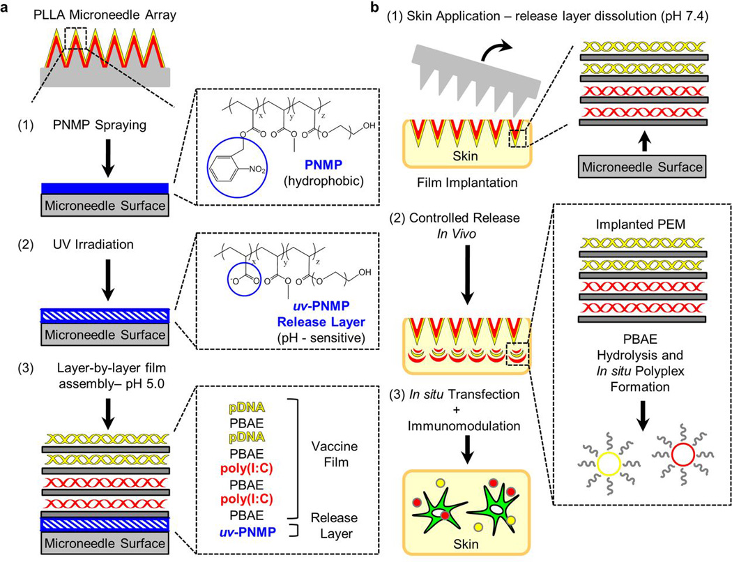
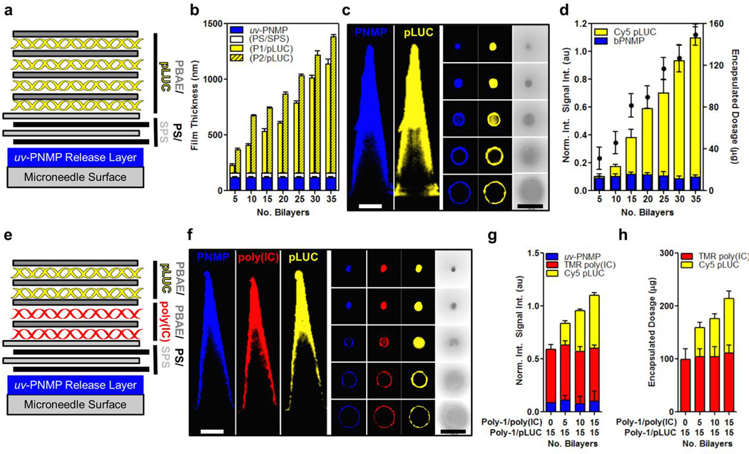
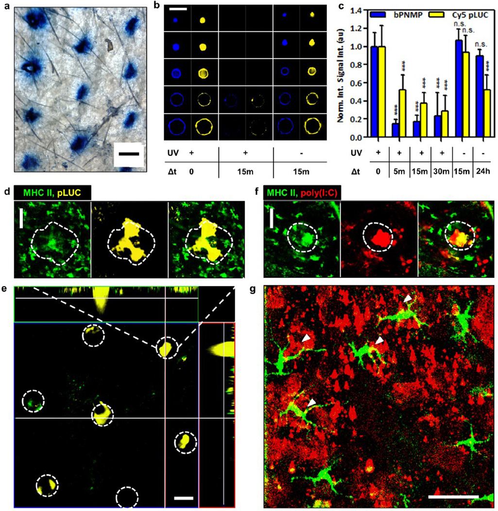
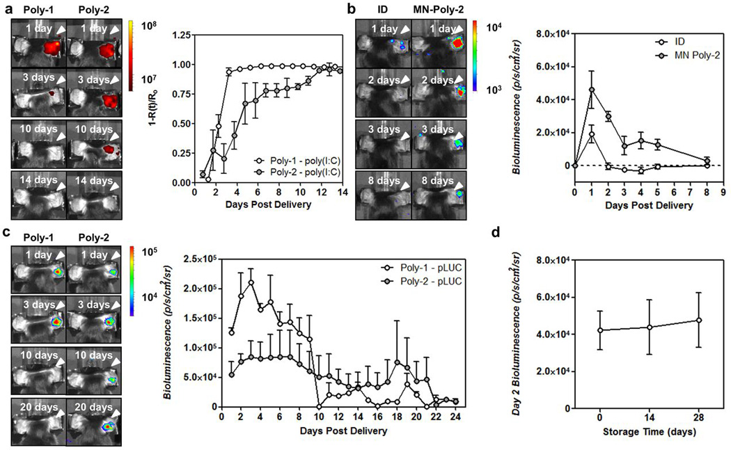
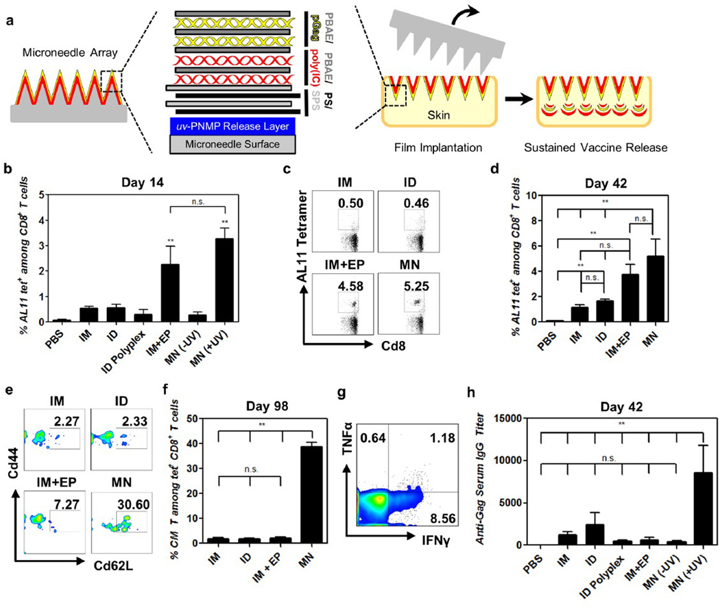
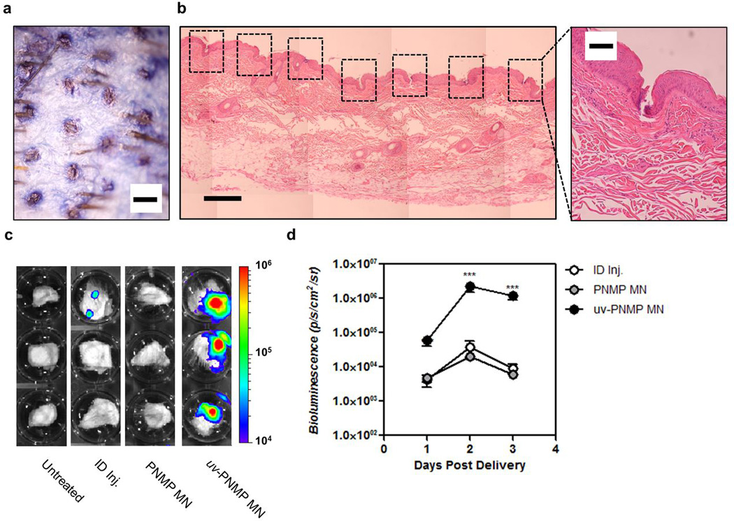
DNA vaccines have many potential benefits but have failed to generate robust immune responses in humans. Recently, methods such as in vivo electroporation have demonstrated improved performance, but an optimal strategy for safe, reproducible, and pain-free DNA vaccination remains elusive. Here we report an approach for rapid implantation of vaccine-loaded polymer films carrying DNA, immune-stimulatory RNA, and biodegradable polycations into the immune-cell-rich epidermis, using microneedles coated with releasable polyelectrolyte multilayers. Films transferred into the skin following brief microneedle application promoted local transfection and controlled the persistence of DNA and adjuvants in the skin from days to weeks, with kinetics determined by the film composition. These 'multilayer tattoo' DNA vaccines induced immune responses against a model HIV antigen comparable to electroporation in mice, enhanced memory T-cell generation, and elicited 140-fold higher gene expression in non-human primate skin than intradermal DNA injection, indicating the potential of this strategy for enhancing DNA vaccination.


Figures

Figure 1
Figure 1. Design of quick-release vaccine-loaded microneedle coatings
a, Schematic view of release-layer-mediated multilayer tattooing strategy using coated microneedles: (1) PLLA microneedles are coated with PNMP release-layer films through spray deposition; (2) UV-irradiation imparts pH-sensitive aqueous solubility to the PNMP film, forming a uv-PNMP ‘release-layer’; (3) Overlying multilayer films containing nucleic acids are constructed using LbL deposition at pH 5.0. b, Mechanism of action for multilayer tattooing: (1) Microneedle application to skin and exposure to interstitial fluid gives rapid release-layer dissolution, mediating overlying film delamination and retention in skin following microneedle removal; (2) Implanted films provide sustained release of nucleic acids through hydrolytic PBAE degradation and release of in situ-formed PBAE/nucleic acid polyplexes; (3) released polyplexes mediate local transfection and immune modulation in the tissue.
Figure 2
Figure 2. LbL assembly of microneedle coatings carrying DNA, immunostimulatory RNA, and transfection agents
a, Film architecture for (uv-PNMP)(PS/SPS)n(PBAE/pLUC)n multilayers. b, Growth of (poly-1/pLUC)n and (poly-2/pLUC)n multilayers assembled onto (uv-PNMP)(PS/SPS)20 films on silicon substrates as a function of the number of deposited (PBAE/pLUC) bilayers as measured by surface profilometry. c, Representative confocal images of PLLA microneedles coated with (SAv488-bPNMP)(PS/SPS)20(poly-1/Cy5-pLUC)35 films (left – transverse optical sections, right – lateral sections, 100µm z-intervals, scale bars 200 µm. (blue – Sav488-uv-bPNMP, yellow – Cy5-pLUC). d, Quantification of Cy5-pLUC and Sav488-bPNMP incorporated into (SAv488-bPNMP)(PS/SPS)20(poly-1/Cy5-pLUC)n films on microneedles through confocal fluorescence intensity analysis (left axis, n = 15) and measurement of total DNA recovered from dissolved films (right axis, n = 3). e, Film architecture for (uv-PNMP)(PS/SPS)20(Poly-1/pLUC)n(Poly-1/poly(I:C))n multilayers. f, Representative confocal images of microneedles coated with (SAv488-uv-bPNMP)(PS/SPS)20(poly-1/TMR-poly(I:C))15(poly-1/Cy5-pLUC)15 films (left – transverse sections, right – lateral sections, 100µm z-interval, scale 200 µm. (blue – Sav488-uv-bPNMP, yellow – Cy5-pLUC, red – TMR-poly(I:C)). g, h, Quantification of Cy5-pLUC, TMR-poly(I:C), and SAv488-bPNMP incorporated into (SAv488-bPNMP)(PS/SPS)20(poly-1/TMR-poly(I:C))n(poly-1/Cy5-pLUC)n films on microneedles through confocal fluorescence intensity analysis (g, n = 15) and measurement of total nucleic acids recovered from dissolved films (h, n= 3).
Figure 3
Figure 3. PNMP release-layers promote rapid implantation of multilayer films at microneedle penetration sites in vivo
a, Optical micrograph of ear skin stained with trypan blue to reveal epidermal penetration following PLLA microneedle application (scale bar 500µm). b, Representative confocal images of (SAv488-bPNMP)(PS/SPS)20(poly-1/Cy5-pLUC)35-coated PLLA microneedles with or without UV sensitization of the PNMP layer (Blue - Sav488-bPNMP; yellow - Cy5-pLUC), before application, or after 15 min application to murine ear skin (lateral sections, 100 µm z-interval, scale bar 200 µm). c, Quantitation of confocal fluorescence intensities (n = 15) showing loss of Sav488-uv-bPNMP and Cy5-pLUC films from coated microneedles upon application to skin, dependent on UV-induced photo-switching of the PNMP layer solubility. ***, p< 0.0001, analyzed by unpaired t-test. d, Representative confocal image of treated murine skin showing film implantation after 15 min (green, MHC II-GFP; yellow, Cy5-pLUC; penetration site outlined, scale bar 100µm). e, x-y/x-z/y-z confocal images showing depth of Cy5-pLUC film deposition after 15 minute microneedle application (green, MHC II-GFP; yellow, Cy5-pLUC; penetration sites outlined, scale bar 200µm). f, Representative confocal image of treated murine skin showing TMR-poly(I:C) film implantation after 15 min microneedle application (green, MHC II-GFP; red, TMR-poly(I:C); penetration site outlined, scale bar 100 µm). g, Colocalization and uptake of TMR-poly(I:C) by MHC II-GFP+ LCs at microneedle insertion site 24 hrs following film implantation (green, MHC II-GFP; red, TMR-poly(I:C); yellow, overlay, scale bar 50 µm).
Figure 4
Figure 4. Implanted films control the physical and functional persistence of pDNA and poly(I:C) in vivo
a, Representative whole-animal fluorescence images showing TMR-poly(I:C) retention at the application site and quantitative analysis of normalized total fluorescence R(t) relative to initial fluorescence Ro from groups of animals (n = 3) over time following 15 min application of PLLA microneedles coated with (uv-PNMP)(PS/SPS)20(PBAE/TMR-poly(I:C))35 multilayers containing poly-1 or poly-2 as the PBAE component. b, Representative whole-animal luminescent images and quantitative analysis of luminol signal from MPO-dependent oxidative burst in activated phagocytes at the treatment site over time following intradermal injection of 10 µg poly(I:C) or 15 min application of PLLA microneedles coated with (uv-PNMP)(PS/SPS)20(Poly-2/poly(I:C))35 multilayers. c, Representative whole animal bioluminescence images of pLUC expression at the application site and mean bioluminescence intensity over time following 15 minute application of microneedles coated with (uv-PNMP)(PS/SPS)20(PBAE/pLUC)35 multilayers containing poly-1 or poly-2 as the PBAE component. d, Mean bioluminescent intensity on day 2 following 15 min application of microneedles coated with (uv-PNMP)(PS/SPS)20(Poly-1/pLUC)35 multilayers stored dry at 25°C for 0, 14, or 28 days.
Figure 5
Figure 5. Microneedle tattooing with multilayer films carrying pDNA and poly(I:C) generates potent cellular and humoral immunity against a model HIV antigen
a, C57Bl/6 mice (n = 4 mice/group) were immunized with 20 µg pGag and 10 µg poly(I:C) on days 0 and 28 intramuscularly (with or without electroporation (EP)) in the quadriceps, intradermally in the dorsal ear skin (with free pGag or pGag/poly-1 polyplexes, ID ± Polyplex), or by 15 minute application of (PNMP)(PS/SPS)20(poly-1/poly(I:C))35(poly-1/pLUC)35–coated microneedles without or without UV priming of the PNMP release-layer (MN ± UV) to the dorsal ear skin. b-d, Frequency of Gag-specific CD8+ T-cells in peripheral blood assessed by flow cytometry analysis of tetramer+ CD8+ T-cells. Shown are mean tetramer+ values from b, day 14 and c, representative cytometry plots from individual mice and d, mean tetramer+ values from day 42. e-f, Analysis of T-cell effector/central memory phenotypes in peripheral blood by CD44/CD62L expression of tetramer+ cells from peripheral blood. Shown are e, representative cytometry plots from individual mice at day 49 and f, mean percentages of tetramer+CD44+CD62L+ among CD8+ T cells at day 98. g, Mice immunized with microneedles were recalled on day 105 by IM injection of 50 µg pGag, and assessed for cytokine production on ex vivo restimulation with AL11 peptide on day 112. Shown is representative flow cytometry analysis of IFN-γ/TNF-α-producing CD8+ T-cells. h, Enzyme-linked-immunosorbent assay analysis of total Gag-specific IgG in sera at day 42. **, p< 0.005, analyzed by two-way ANOVA.
Figure 6
Figure 6. Multilayer tattooing enhances transfection in non-human primate skin
a, Optical micrograph of macaque quadriceps skin showing microneedle penetration pattern stained using trypan blue (scale bar 500µm). b, Histological section of microneedle-treated macaque skin showing epidermal disruption at microneedle insertion sites (boxed, left, scale bar 500µm; right, scale bar 100µm). c, Bioluminescence images of luciferase expression 2 days following pLUC delivery by ID injection or microneedle tattooing with (PS/SPS)20(poly-1/pLUC)35 films from either uv-PNMP- or non-irradiated PNMP-coated microneedles following a 15 minute application. d, Quantification of total bioluminescent signal in cultured skin tissue explants 1, 2, and 3 days following treatment. ***, p< 0.0001, analyzed by unpaired t-test.

References

    1. Kutzler MA, Weiner DB. DNA vaccines: ready for prime time? Nat. Rev. Genet. 2008;9:776–788. - PMC - PubMed
    1. Rice J, Ottensmeier CH, Stevenson FK. DNA vaccines: precision tools for activating effective immunity against cancer. Nat. Rev. Cancer. 2008;8:108–120. - PubMed
    1. Ulmer JB, Wahren B, Liu MA. Gene-based vaccines: Recent technical and clinical advances. Trends Mol. Med. 2006;12:216–222. - PubMed
    1. Sardesai NY, Weiner DB. Electroporation delivery of DNA vaccines: prospects for success. Curr. Opin. Immunol. 2011;23:421–429. - PMC - PubMed
    1. Weir E, Hatch K. Preventing cold chain failure: vaccine storage and handling. CMAJ. 2004;171:1050. - PMC - PubMed

Publication types