Functions and imaging of mast cell and neural axis of the gut
- PMID: 23354018
- PMCID: PMC3922647
- DOI: 10.1053/j.gastro.2013.01.040
Functions and imaging of mast cell and neural axis of the gut
Abstract
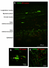
Close association between nerves and mast cells in the gut wall provides the microanatomic basis for functional interactions between these elements, supporting the hypothesis that a mast cell-nerve axis influences gut functions in health and disease. Advanced morphology and imaging techniques are now available to assess structural and functional relationships of the mast cell-nerve axis in human gut tissues. Morphologic techniques including co-labeling of mast cells and nerves serve to evaluate changes in their densities and anatomic proximity. Calcium (Ca(++)) and potentiometric dye imaging provide novel insights into functions such as mast cell-nerve signaling in the human gut tissues. Such imaging promises to reveal new ionic or molecular targets to normalize nerve sensitization induced by mast cell hyperactivity or mast cell sensitization by neurogenic inflammatory pathways. These targets include proteinase-activated receptor (PAR) 1 or histamine receptors. In patients, optical imaging in the gut in vivo has the potential to identify neural structures and inflammation in vivo. The latter has some risks and potential of sampling error with a single biopsy. Techniques that image nerve fibers in the retina without the need for contrast agents (optical coherence tomography and full-field optical coherence microscopy) may be applied to study submucous neural plexus. Moreover, the combination of submucosal dissection, use of a fluorescent marker, and endoscopic confocal microscopy provides detailed imaging of myenteric neurons and smooth muscle cells in the muscularis propria. Studies of motility and functional gastrointestinal disorders would be feasible without the need for full-thickness biopsy.
Copyright © 2013 AGA Institute. Published by Elsevier Inc. All rights reserved.
Figures


References
-
- Buhner S, Schemann M. Mast cell-nerve axis with a focus on the human gut. Biochim Biophys Acta. 2012;1822:85–92. - PubMed
-
- Bischoff SC. Physiological and pathophysiological functions of intestinal mast cells. Semin Immunopathol. 2009;31:185–205. - PubMed
-
- Lorentz A, Bischoff SC. Regulation of human intestinal mast cells by stem cell factor and IL-4. Immunol Rev. 2001;179:57–60. - PubMed
-
- Aldenborg F, Enerbäck L. The immunohistochemical demonstration of chymase and tryptase in human intestinal mast cells. Histochem J. 1994;26:587–596. - PubMed
Publication types
MeSH terms
Grants and funding
LinkOut - more resources
Full Text Sources
Other Literature Sources
Medical

