. 2013 Mar;15(3):249-60.
doi: 10.1038/ncb2679. Epub 2013 Jan 27.

Spatial regulation of VEGF receptor endocytosis in angiogenesis

Affiliations

Spatial regulation of VEGF receptor endocytosis in angiogenesis

Masanori Nakayama et al. Nat Cell Biol. 2013 Mar.

Abstract

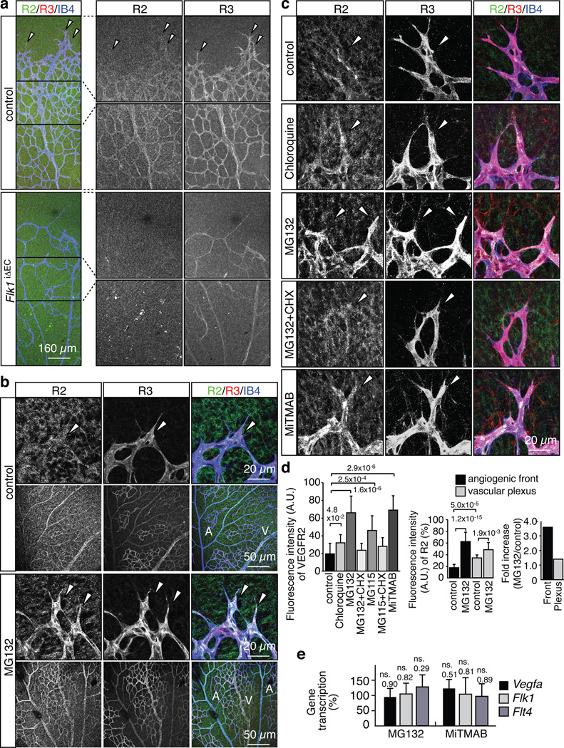
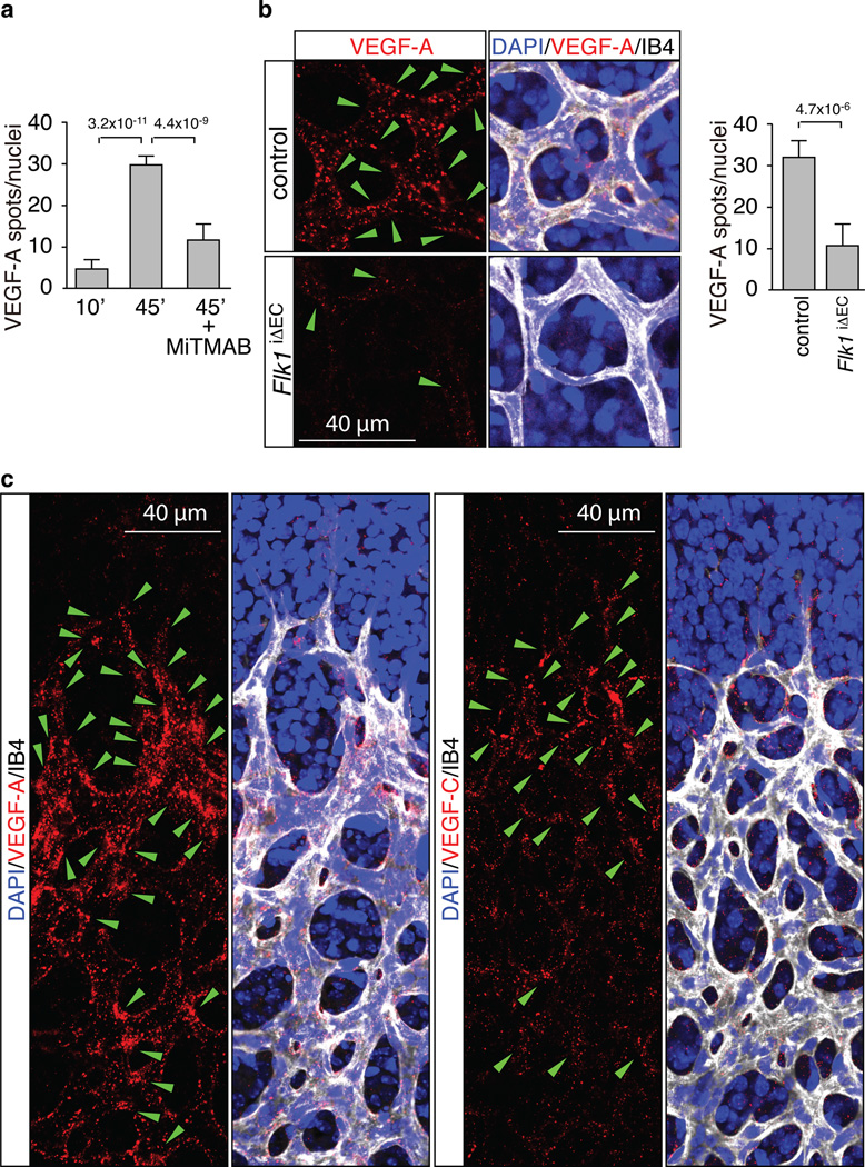
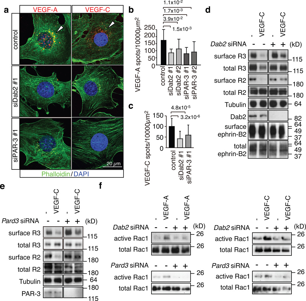
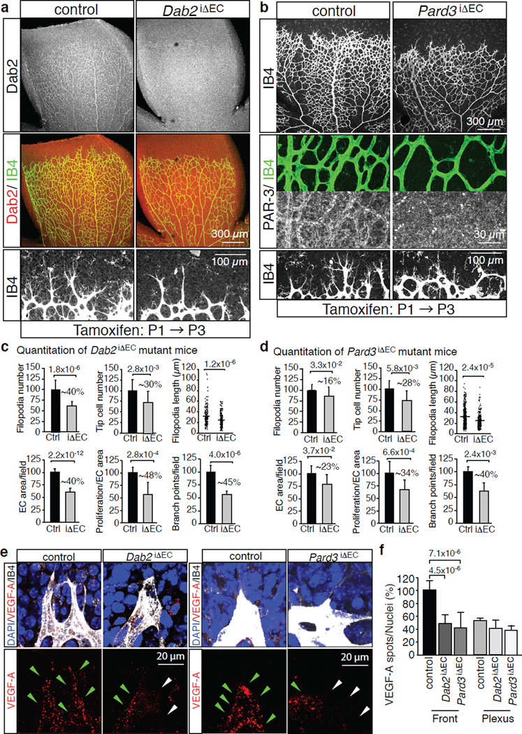
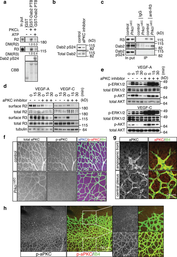
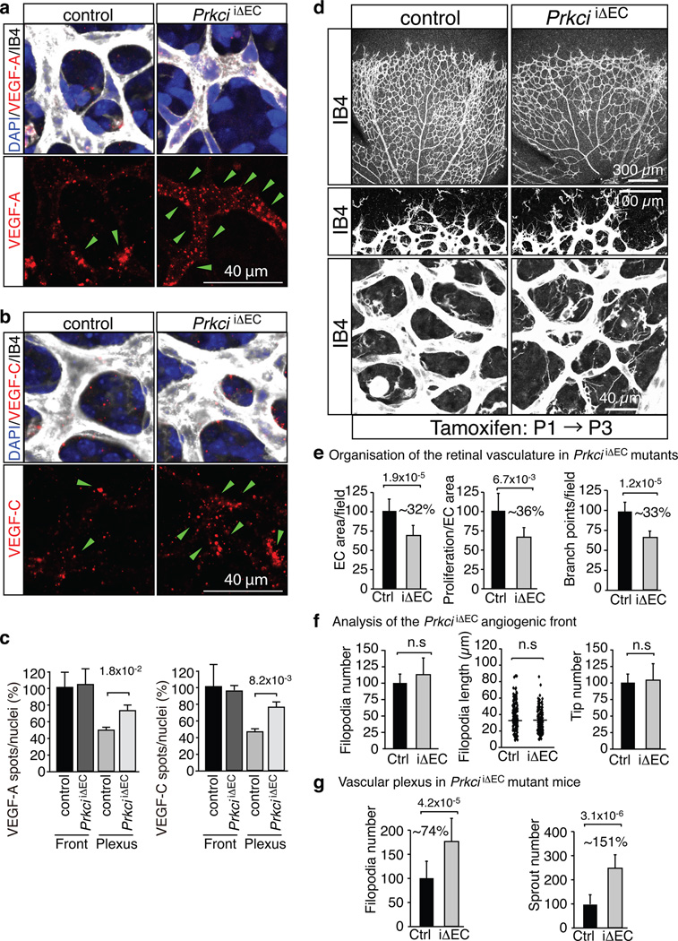
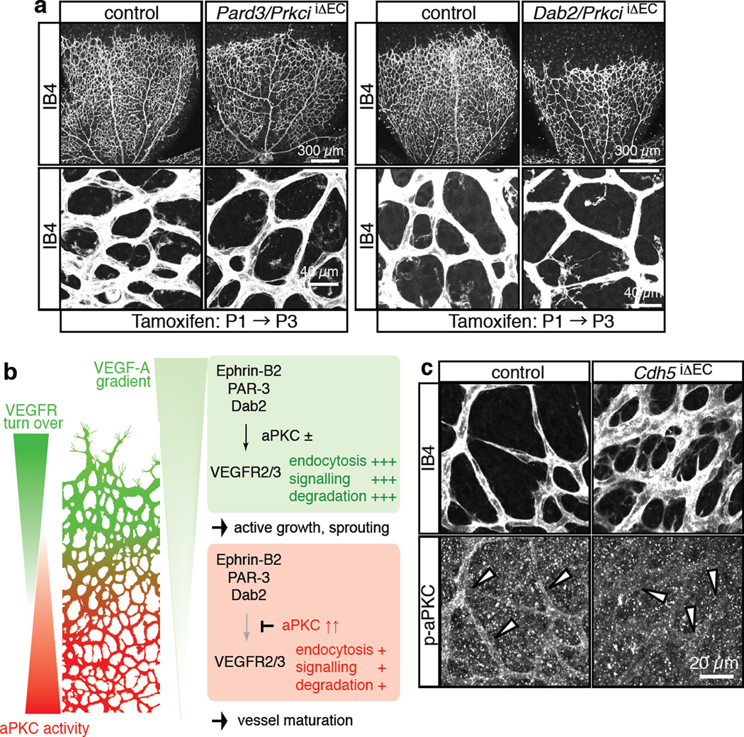
Activities as diverse as migration, proliferation and patterning occur simultaneously and in a coordinated fashion during tissue morphogenesis. In the growing vasculature, the formation of motile, invasive and filopodia-carrying endothelial sprouts is balanced with the stabilization of blood-transporting vessels. Here, we show that sprouting endothelial cells in the retina have high rates of VEGF uptake, VEGF receptor endocytosis and turnover. These internalization processes are opposed by atypical protein kinase C activity in more stable and mature vessels. aPKC phosphorylates Dab2, a clathrin-associated sorting protein that, together with the transmembrane protein ephrin-B2 and the cell polarity regulator PAR-3, enables VEGF receptor endocytosis and downstream signal transduction. Accordingly, VEGF receptor internalization and the angiogenic growth of vascular beds are defective in loss-of-function mice lacking key components of this regulatory pathway. Our work uncovers how vessel growth is dynamically controlled by local VEGF receptor endocytosis and the activity of cell polarity proteins.


Figures

Figure 1
Figure 1. High VEGF receptor turnover in the angiogenic front
a, Anti-VEGFR2 (green), VEGFR3 (red) and IsolectinB4 (IB4; blue) staining of the P6 retinal vasculature. Absent endothelial VEGFR2 immunostaining and strongly decreased VEGFR3 in Flk1iΔEC retinas. Arrowheads indicate distal sprouts. b, Strong upregulation of VEGFR2 (green) and VEGFR3 (red) protein levels in P6 vessel sprouts (arrowheads) compared to vessels in central retina at 2 hours after intraocular MG132 injection. ECs, Isolectin B4 (IB4, blue). Arteries (A) and veins (V) are indicated. c, VEGFR2 and VEGFR3 immunostaining at 2 hours after intraocular injection of the indicated inhibitors. ECs, Isolectin B4 (IB4, blue). Arrowheads mark sprouts. d, Statistical analysis of VEGFR2 staining as shown in (b) and (c). Data represent the means±s.d. of 12 independent experiments. P values, ANOVA. e, qPCR analysis showing that Vegfa, Vegfr2/Flk1 and Vegfr3/Flt4 transcript levels in eyes were not significantly increased after MG132 or MiTMAB treatment. Data represent the means±s.d. of 6 independent experiments. P values, two-tailed Student’s t-test compared to vehicle-injected controls. ns, not significant.
Figure 2
Figure 2. Spatial differences in VEGF uptake in the retina
a, Quantitation of Alexa-labelled VEGF-A in retinal ECs at 10 and 45 min after injection. The formation of intracellular spots was blocked by MiTMAB. Data represent the means±s.d. of 6 independent experiments. P values, ANOVA. b, VEGF-A uptake in blood vessels of FlkiΔEC mice. Green arrowheads indicate internalised Alexa dye-coupled VEGF-A (red). Cell nuclei, DAPI (blue); ECs, Isolectin B4 (IB4, white). Data represent the means±s.d. of 6 independent experiments. P values, two-tailed student t test. c, Spatial distribution of VEGF-A or VEGF-C (red) uptake in retinal vessels. Green arrowheads indicate internalised label spots. Cell nuclei, DAPI (blue); ECs, Isolectin B4 (IB4, white).
Figure 3
Figure 3. Identification of Dab2 and PAR-3 as interactors of ephrin-B2 and VEGF receptors
a, Domain structure and deletion constructs of ephrin-B2, Dab2 and PAR-3. Numbers refer to amino-acid positions in the murine gene products. TM, transmembrane region; CR1, conserved region 1; CR3, conserved region 3; PDZ, PSD-95/Dlg/ZO-1 domains; PTB, phosphotyrosine binding region. b, Endocytic proteins identified by mass spectrometry. Name of proteins and corresponding genes, the number of peptides identified, and PEP scores are listed. c, Immunoblotting of affinity-purified samples with anti-Dab2 antibody. Pull-down was performed with GST-cyto WT (GST-fusion protein of cytoplasmic region of ephrin-B2) or GST-cyto VS (mutation in the C-terminal PDZ binding motif of ephrin-B2). Molecular weight marker (kD) is indicated on the right. d, e, Pull-down analysis with the indicated domains of PAR-3 (c) or Dab2 (d) fused to GST. Interacting VEGFR2, VEGFR3, Dab2 and ephrin-B2 from cultured mouse ECs were detected by immunoblot. Binding of VEGF receptors to the GST-PTB domain of Dab2 was enhanced by VEGF. Asterisks mark bait proteins in Coomassie Brilliant Blue (CBB) staining (bottom panels). f, Immunoprecipitation (IP) of endogenous Dab2 and PAR-3 with VEGFR3 from lung lysate of control mice but not Pard3iΔEC mutants. g, Association of PAR-3, clathrin heavy chain (CHC) and VEGFR3 with immunoprecipitated (IP) VEGFR2 in VEGF-C-treated mouse ECs. CHC interaction with VEGFR2 was reduced after knockdown of Dab2 (siDab2).
Figure 4
Figure 4. Dab2 and PAR-3 control VEGF receptor internalisation
a, Alexa546-labelled VEGF-A or VEGF-C (red) accumulated in the perinuclear region of control mouse ECs at 30 min after stimulation, which was strongly reduced after knockdown of Dab2 or Pard3. Actin, Phalloidin (green); nuclei, DAPI (blue). b, c, Quantitation of Alexa546-positive peri-nuclear VEGF-A (b) or VEGF-C (c) spots. Two different siRNAs were used for Dab2 and Pard3 in (b). Data represent the means±s.d. of 6 independent experiments. P values, two-tailed Student’s t-test. At least 100 cells were scored in each experiment. d, e, Biochemical detection of biotinylated (surface) VEGFR2 and VEGFR3 in stimulated control and Dab2 (d) or Pard3 (e) KD cells. Antibodies used for immunoblotting and molecular weight marker are indicated. f, Activation of Rac1 in control and Dab2 or Pard3 KD mouse ECs stimulated with VEGF-A or VEGF-C for 5 min, as indicated.
Figure 5
Figure 5. Endothelial Dab2 and PAR-3 regulate angiogenic vessel growth
a, Overview of the P6 control and Dab2iΔEC retinal vasculature. Anti-Dab2 (white/red) staining is shown in top panels. ECs, Isolectin B4 (IB4). Bottom panels show higher magnification of the angiogenic front. b, Defects in the P6 Pard3iΔEC retinal vasculature. Anti-PAR-3 and IB4 (green) staining in middle panels show successful deletion of PAR-3 in Pard3iΔEC vessels. Residual round signals correspond to autofluorescent blood cells. Bottom panels show higher magnification of the angiogenic front. c, d, Quantitation of filopodia number and length, tip cell number, EC-covered area, EC proliferation and vessels branch points in Dab2 mutant (iΔEC) (c) or Pard3 mutant (d) retinas compared to the corresponding control littermates (Ctrl). Dab2 mutant n=7, Pard3 mutant n=3. Percentage of reduction is indicated. Data represent the means±s.d. P values, two-tailed Student’s t-test. e, Reduced uptake of labelled VEGF-A (red) at the Dab2iΔEC or Pard3iΔEC angiogenic front compared to control littermates. Green arrowheads indicate VEGF-A spots, white arrowheads marks ECs with no or little uptake. f, Statistical analysis of internalised Alexa-coupled VEGF-A in the angiogenic front and central plexus. Data represent the means±s.d. of 6 independent experiments. P values, two-tailed Student’s t-test.
Figure 6
Figure 6. Negative regulation of VEGF receptor internalisation by aPKC
a, Phosphorylation of a GST-Dab2 fusion protein by recombinant PKCλ reduced its interaction with VEGFR3 and VEGFR2 in pull down assays. Densiometric (DM) values are shown below bands. ATP for the kinase activation was added (+) or absent (−), as indicated. CBB, Coomassie Brilliant Blue staining of GST fusion proteins. b, Western blot showing reduced Dab2 phoshorylation at Serine 24 (pS24) in cultured ECs after 30 min incubation with 5µM aPKC inhibitor. c, Association of Dab2 with immunoprecipitated VEGFR3 (anti-R3) was enhanced in PrkciiΔEC lung lysate compared to control littermates. No specific bands were immunoprecipitated with IgG. VEGFR3-associated Dab2 lacked detectable phosphorylation in Ser24 (pS24). d, Effect of aPKC inhibition on VEGF-A- or VEGF-C-induced VEGF receptor internalisation at indicated time points. Cells were preincubated with 5 µM of aPKC inhibitor for 30 min. Surface VEGFR2 (R2) and VEGFR3 (R3) were identified by biotinylation. e, Increased activation of MAP kinase (p-ERK1/2) by VEGF-A or VEGF-C after aPKC inhibition in cultured mouse ECs. Effects on AKT phosphorylation were comparably modest. Total ERK1/2 and AKT are shown as loading controls. Molecular weight marker (kD) is indicated. f, Anti-aPKC (total), anti-phospho-aPKC (p-aPKC, Thr560) and Isolectin B4 (IB4) staining of the P6 control and PrkciiΔEC retinal vasculature. g, Anti-aPKC immunostaining (red) labels ECs (IB4, green) at the angiogenic front and in the central retinal plexus. h, Phospho-aPKC (p-aPKC, Thr560) immunosignals were weak at the angiogenic front in comparison to vessels of the central plexus. Higher magnification of p-aPKC signals and merged channels in insets is shown on the right.
Figure 7
Figure 7. Increased VEGF uptake and sprouting in the PrkciiΔEC central retina
a, b, VEGF-A (red, a) or VEGF-C (b) uptake in the PrkciiΔEC central plexus. Green arrowheads indicate ligand spots. Cell nuclei, DAPI (blue); ECs, Isolectin B4 (IB4, white). c, Statistical analysis of internalised VEGFs as shown in (a) and (b). Data represent the means±s.d. of 6 independent experiments. P values, two-tailed Student’s t-test. d, Phenotype of the Isolectin B4-stained (IB4) P6 PrkciiΔEC retinal vasculature. Higher magnification of the angiogenic front (middle) and central plexus (bottom) are shown. e–g, Quantitation of vessels branch points, EC area and proliferation (e, n=3), the number and length of filopodia and the number of distal sprout tips at the angiogenic front (f, n=6), and the number of ectopic sprouts and filopodia (g, n=6) in the central retina of Prkci mutants (iΔEC) compared to control (Ctrl) retinas. Data represent the means±s.d. P values, two-tailed Student’s t-test
Figure 8
Figure 8. Schematic depiction of key findings
a, Absence of ectopic sprouting and filopodia in the central retina of Pard3/PrkciiΔEC (left) or Dab2/PrkciiΔEC (right) double mutant mice (compare to lower panels to equivalent regions shown in Fig. 7d). ECs, Isolectin B4 (IB4, white). b, VEGF receptor turnover (i.e. internalisation, degradation and new synthesis) is highest in vessel sprouts at the angiogenic front compared to the more mature central vessel plexus. This behaviour is not solely controlled by the VEGF-A gradient, but also intrinsic properties of ECs. VEGF receptor internalisation and certain downstream signalling processes are controlled by the transmembrane protein ephrin-B2 and its interaction partners PAR-3 and Dab2. Abundant active (phosphorylated) aPKC in the central retina antagonizes VEGF receptor internalisation by phosphorylating the Dab2 (cargo-binding) PTB domain. This contributes to regional differences in VEGF receptor turnover and VEGF ligand internalisation. We propose that regionally distinct behaviours of ECs in the growing vasculature are modulated by spatially regulated endocytosis. c, Anti-phospho-aPKC (p-aPKC, Thr403; Cell Signaling, lot 8) and Isolectin B4 (IB4) staining of P6 control and Cdh5iΔEC retinal vessels.

Comment in

References

    1. Palamidessi A, et al. Endocytic trafficking of Rac is required for the spatial restriction of signaling in cell migration. Cell. 2008;134:135–147. - PubMed
    1. Le Roy C, Wrana JL. Clathrin- and non-clathrin-mediated endocytic regulation of cell signalling. Nat. Rev. Mol. Cell Biol. 2005;6:112–126. - PubMed
    1. Bush JO, Soriano P. Ephrin-B1 forward signaling regulates craniofacial morphogenesis by controlling cell proliferation across Eph-ephrin boundaries. Genes Dev. 2010;24:2068–2080. - PMC - PubMed
    1. Bryant DM, Mostov KE. From cells to organs: building polarized tissue. Nat. Rev. Mol. Cell Biol. 2008;9:887–901. - PMC - PubMed
    1. Olsson AK, Dimberg A, Kreuger J, Claesson-Welsh L. VEGF receptor signalling - in control of vascular function. Nat. Rev. Mol. Cell Biol. 2006;7:359–371. - PubMed

Publication types

MeSH terms