. 2013 Mar;16(3):281-9.
doi: 10.1038/nn.3319. Epub 2013 Jan 27.

Epigenetic control of female puberty

Affiliations

Epigenetic control of female puberty

Alejandro Lomniczi et al. Nat Neurosci. 2013 Mar.

Abstract

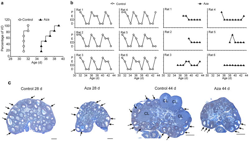
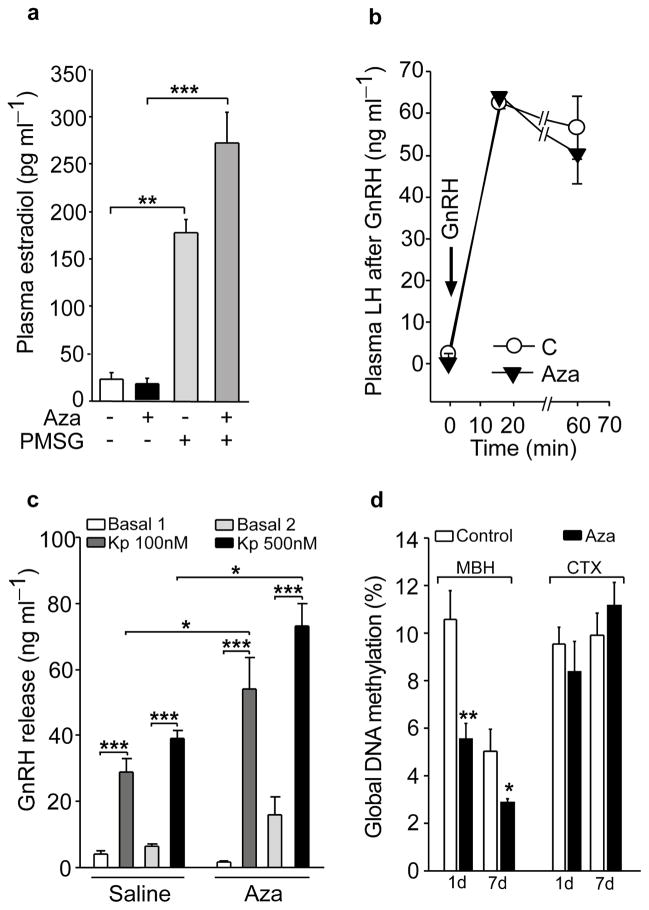
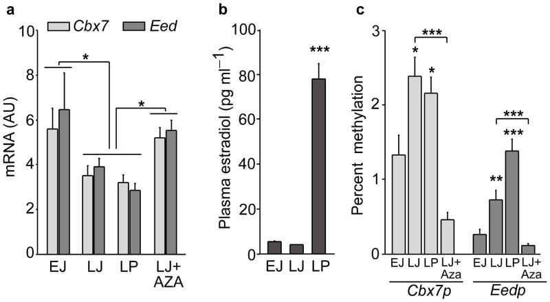
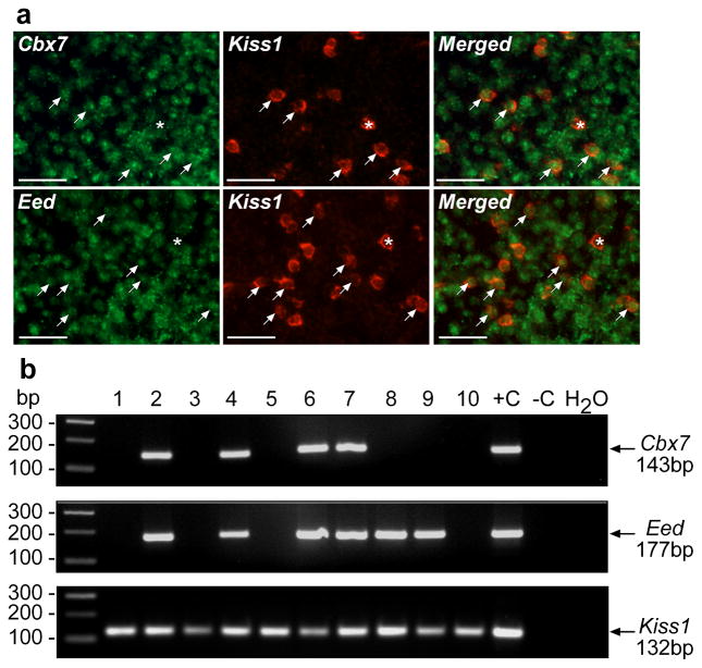
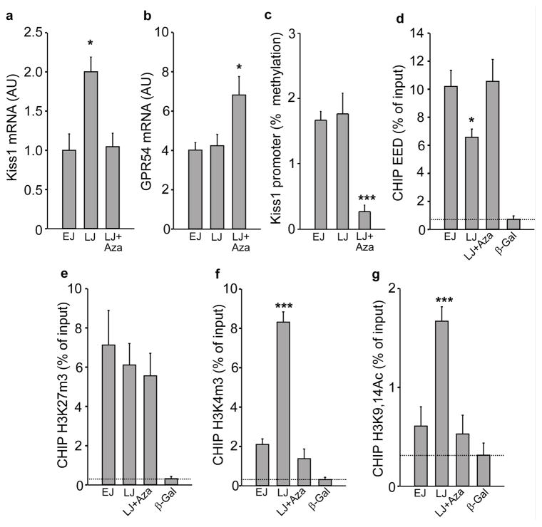
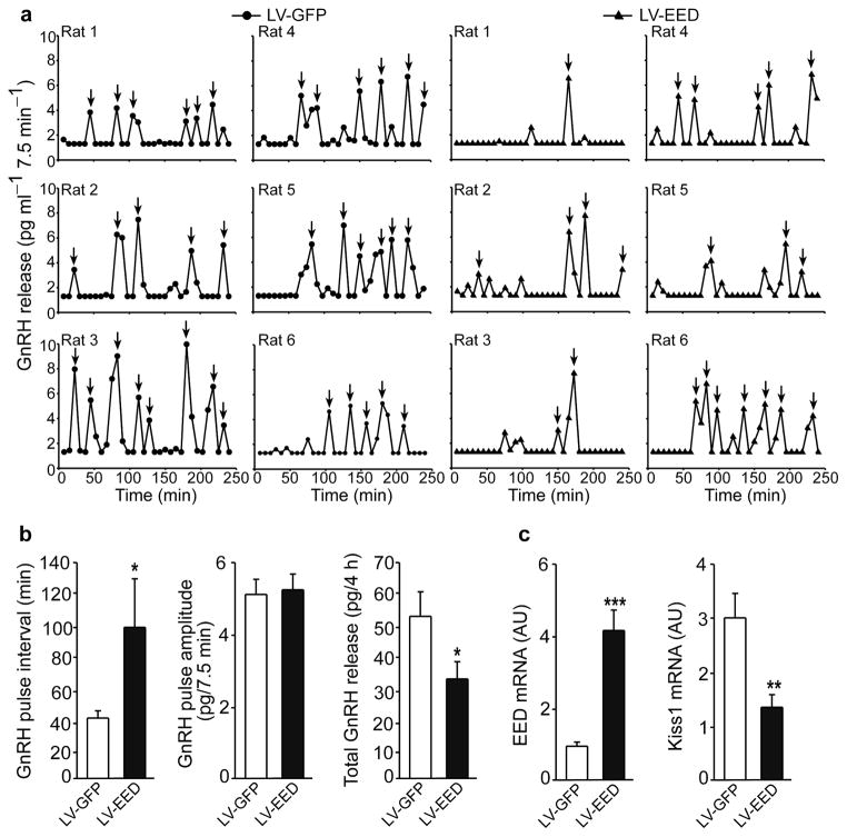
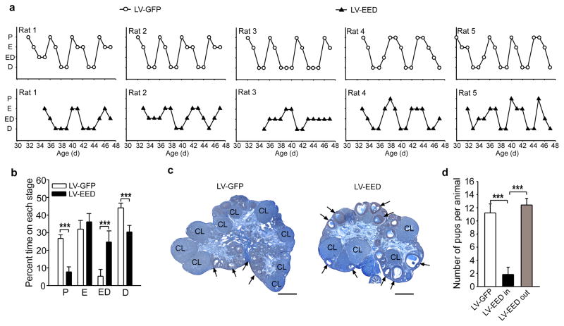
The timing of puberty is controlled by many genes. The elements coordinating this process have not, however, been identified. Here we show that an epigenetic mechanism of transcriptional repression times the initiation of female puberty in rats. We identify silencers of the Polycomb group (PcG) as principal contributors to this mechanism and show that PcG proteins repress Kiss1, a puberty-activating gene. Hypothalamic expression of two key PcG genes, Eed and Cbx7, decreased and methylation of their promoters increased before puberty. Inhibiting DNA methylation blocked both events and resulted in pubertal failure. The pubertal increase in Kiss1 expression was accompanied by EED loss from the Kiss1 promoter and enrichment of histone H3 modifications associated with gene activation. Preventing the eviction of EED from the Kiss1 promoter disrupted pulsatile gonadotropin-releasing hormone release, delayed puberty and compromised fecundity. Our results identify epigenetic silencing as a mechanism underlying the neuroendocrine control of female puberty.


Conflict of interest statement

COMPETING FINANCIAL INTERESTS

The authors declare no competing financial interests.

Figures

Figure 1
Figure 1. In vivo inhibition of DNA methylation results in pubertal failure
(a) Female rats treated with 5-Azacytidine (Aza; i.p., 2mg/kg BW/day) from PND22 onwards have delayed vaginal opening. At the time when vaginal opening has occurred in all control animals (PND32), vaginal patency was not apparent in any of the Aza-treated rats. (b) Estrous cycle profiles showing that Aza treatment markedly disrupts estrous cyclicity. The phases of the estrous cycle depicted on the Y axis are proestrous (P), estrous (E), a transitional stage estrous-diestrous (ED), and diestrous (D). (c) Microphotographs illustrating the delay in ovarian maturation caused by Aza treatment during the juvenile phase of prepubertal development (PND 22 to 28) and the absence of ovulation, assessed by the lack of corpora lutea (CL) in ovaries from young adult (PND day 44) rats. Arrows point to examples of antral follicles. Scale bars = 100 μm.
Figure 2
Figure 2. In vivo inhibition of DNA methylation prevents puberty by disrupting developmental events upstream from the GnRH-pituitary-ovarian axis
(a) Treatment with Aza (2mg/kg BW/day/7days, PND 22–28) does not prevent, but instead augments, the estradiol response of the ovary to stimulation with gonadotropins (PMSG, 8 IU/rat, given as a single i.p injection on PND 26; two days before blood collection)(F3,31=47.81, p<0.001, One-Way ANOVA followed by the SNK multiple comparison test, n=8 animals/group). (b) GnRH (6.5 μg/kg i.p) injection increases plasma LH similarly in control and Aza treated animals (there is a statistically significant difference between time points within each treatment, F2,7=277.23, p<0.001, but not between treatments, F1,7=0.46, p=0.52 with no significant interaction between time points and treatments, F2,47=0.56, p=0.58, Two Way Repeated Measures ANOVA, n=8 rats/group). (c) Hyper-response of the GnRH system to kisspeptin, as determined by the in vitro release of GnRH from ME-ARC fragments (derived from PND 28 rats) incubated with either 100 or 500 nM of Kisspeptin-10 (Kp) (there is a statistically significant interaction between treatment group and Kp dose, F3,47=5.93, p=0.007, Two Way Repeated Measures ANOVA, n=6 rats/group). (d) The Aza treatment reduces global DNA methylation in the female rat hypothalamus but not in the cerebral cortex (CTX) as compared with diluent-treated controls (C) (In MBH: One day of treatment t=3.65, p=0.004, 7 days of treatment t=2.98, p=0.044. In CTX no statistical significance was found, t=0.803, p=0.441 and t=−0.928, p=0.375 one and seven day treatment respectively, Student t Test, n=6 rats/group). Columns or circles are means and vertical lines represent S.E.M. *p<0.05; **p<0.01; ***p<0.001
Figure 3
Figure 3. The initiation of puberty is accompanied by increased promoter methylation and decreased expression of two PcG genes required for PcG-mediated gene silencing in the medial basal hypothalamus (MBH)
(a) Cbx7 and Eed mRNA expression decrease in the MBH at the initiation of puberty and this decrease is prevented by treatment with Aza. EJ = early juvenile; LJ = late juvenile; LP = late proestrus, the day of the first preovulatory surge of gonadotropins. (For Cbx7 expression: F3,31= 3.72, p=0.022 and for Eed expression: F3,31= 6.07, p=0.003, One Way ANOVA, n=6–10 rats per group). (b) Changes in plasma estradiol levels at the time of female puberty (F2,17=109.18, p<0.001, One Way ANOVA, n=6 rats/group). (c) Methylation of the promoter regions of Cbx7 and Eed (Cbx7p and Eedp, respectively) increases in the MBH with the advent of puberty as determined by mCytosine immunoprecipitation followed by real time PCR of immunoprecipitated DNA. Treatment with Aza prevents the increase in promoter methylation. The results are expressed as percent methylation (signal from mC immunoprecipitated DNA/signal from input DNA x 100) (Cbx7p methylation: F3,18=17.6, p<0.001 and Eedp methylation: F3,18=23.22, p<0.001, One Way ANOVA, n=4–5 rats per group). Columns are means and vertical bars are S.E.M. *p<0.05; **p<0.01; ***p<0.001 vs. EJ or Aza groups (in all cases ANOVA was followed by the SNK multiple comparison test for unequal replications).
Figure 4
Figure 4. The Cbx7 and Eed genes are expressed in kisspeptin neurons of the ARC
(a) Double fluorescent in situ hybridization showing the presence of Cbx7 and Eed mRNA transcripts (green color) in Kiss1 mRNA containing neurons (red color). Arrows point to double positive neurons; asterisks denote Kiss1-mRNA positive neurons without detectable Cbx7 or Eed mRNA. Scale bar= 20μm. (b) Single cell-PCR of eGFP-tagged kisspeptin neurons demonstrating the presence of Cbx7 and Eed mRNAs in these cells.
Figure 5
Figure 5. Increased Kiss1 expression in the MBH at the initiation of puberty is accompanied by eviction of EED from the Kiss1 promoter and changes in associated repressive and activating histone modifications, without changes in DNA methylation
(a) Kiss1 mRNA abundance increases in the MBH at LJ and this increase is prevented by inhibition of DNA methylation (F2,21=7.11, p=0.005),. (b) Gpr54 mRNA abundance does not change in the MBH at the onset of puberty, but increases after inhibition of DNA methylation (F2,21=4.78, p=0.021). (c) Methylation of the Kiss1 promoter in the MBH does not change at puberty but it is diminished by inhibition of DNA methylation (F2,13=13.93, p<0.001). (d) EED is evicted from the Kiss1 promoter at the initiation of puberty and this eviction fail to occur in Aza treated animals (F3,19=17.98, p<0.001). (e) The association of H3K27m3 to the Kiss1 promoter does not decrease at LJ and is not affected by Aza (F3,22=3.45, p=0.037). (f) The abundance of H3K4m3 increases at LJ and the increase is prevented by inhibition of de novo DNA methylation (F3,23=76.09, p<0.001). (g) The association of H3K9,14Ac also increases at LJ and this increase fails to occur in Aza treated rats (F3,23=12.43, p<0.001). For all panels n=6–8 animals/group, except panel c (n=4–5). Columns are means and vertical bars are S.E.M. *p<0.05; ***p<0.001 vs. EJ (in all cases One Way ANOVA was followed by the SNK multiple comparison test for unequal replications). Antibodies to β–Galactosidase (β–Gal) (a protein not expressed in the rat) was used as negative control; dotted line depicts minimum sensitivity level of the technique.
Figure 6
Figure 6. EED delivered to the ARC of immature female rats is recruited to the Kiss1 promoter and represses kisspeptin expression
Double immunostaining for GFP (Green) and Kisspeptin (Red) in the ARC of an LV-EED injected 44 day-old rat. (a) Cell nuclei of the ARC region identified by DAPI staining (blue) and transduction of ARC neurons by the LV-EED construct. (b) Kisspeptin positive neurons transduced by LV-EED. (c) Higher magnification view of a kisspeptin neuron transduced with the LV-EED construct. Scale bars: 100μm (a–b), 10μm (c). (d) CHIP assay from two controls (LV-GFP) and two experimental (LV-EED) animals showing association of EED-HA to a genomic region that includes the Kiss1 transcription start site as determined by PCR amplification of DNA immunoprecipitated with antibodies recognizing the HA epitope tagging EED. (e) Detection of kisspeptin neuron cell bodies in the ARC after micro-injection of either LV-GFP or LV-EED. Red circles identify kisspeptin positive cell bodies.
Figure 7
Figure 7. EED delivered to the ARC of immature female rats inhibits GnRH pulse frequency without changing pulse amplitude
Seven days after stereotaxic delivery of lentiviral particles, ARC-ME explants derived from animals expressing GFP (LV-GFP, circles) or EED (LV-EED, triangles) were incubated for 4h in Krebs-Ringer-Bicarbonate buffer with the medium changed every 7.5min for assessment of pulsatile GnRH release. (a) Individual profiles of GnRH release from LV-GFP and LV-EED injected rats, n = 6 rats/group. Arrows indicate individual pulses. (b) Left panel: GnRH pulse frequency was significantly (t=25.5, p=0.026, Mann-Whitney Rank Sum test) reduced in LV-EED injected animals, as determined by an increase in interpulse interval; Middle panel: pulse amplitude remained unchanged (t=-0.307, p=0.765, Student t Test); Right panel: Total GnRH output/4h incubation was significantly (t=2.61, p=0.026, Student t test) reduced in the LV-EED treated animals. (c) Eed mRNA content was 4-fold higher (t=−5.77, p<0.001) and Kiss1 mRNA content 2 times lower (t=3.17, p=0.01) in LV-EED injected animals than in LV-GFP injected rats (Student t test, in all cases n=6 rats/group). Columns are means and vertical bars are S.E.M..
Figure 8
Figure 8. EED delivered to the ARC of immature female rats delays puberty, disrupts estrous cycle and impairs fertility
(a) Estrous cycles of LV-GFP (circles) and LV-EED (triangles) injected animals. (b) Disruption of estrous cyclicity by LV-EED. Results are shown as percent time spent at each particular phase during a 21-day period. P = proestrous (t=11.96, p<0.001), E = estrous (t=−1.52, p=0.159), ED = transitional stage estrous-diestrous (t=−6.077, p<0.001), D = diestrous (t=7.203, p<0.001) (Student t Test, n= 5 rats/group). (c) Example of ovaries from LV-GFP and LV-EED injected rats collected on PND44. CL indicates corpora luea and arrows point to examples of antral follicles. Scale bars = 100μm. (d) Fertility rate measured as the number of pups delivered after exposing LV-GFP and LV-EED injected females to a fertile male. LV-EED in, injections correctly placed into the ARC; LV-EED out, misplaced injections located outside the ARC. (F2,14=23.92, p<0.001, One Way ANOVA. n=5 rats/group). Columns are means and vertical bars are S.E.M. ***p<0.001 (ANOVA was followed by the SNK multiple comparison test for unequal replications to compare more than two groups).

Comment in

  • A piece in the puzzle of puberty.
    McCarthy MM. McCarthy MM. Nat Neurosci. 2013 Mar;16(3):251-3. doi: 10.1038/nn.3339. Nat Neurosci. 2013. PMID: 23434974 No abstract available.

References

    1. Plant TM, Witchel SF. Puberty in nonhuman primates and humans. In: Neill JD, editor. The Physiology of Reproduction. 3. Academic Press/Elsevier; San Diego: 2006. pp. 2177–2230.
    1. Ojeda SR, Skinner MK. Puberty in the rat. In: Neill JD, editor. The Physiology of Reproduction. 3. Academic Press/Elsevier; San Diego: 2006. pp. 2061–2126.
    1. Kordon C, Drouva SV, Martínez de la Escalera G, Weiner RI. Role of classic and peptide neuromediators in the neuroendocrine regulation of luteinizing hormone and prolactin. In: Knobil E, Neill JD, editors. The Physiology of Reproduction. 2. Vol. 1. Raven Press; New York: 1994. pp. 1621–1681.
    1. Ojeda SR, Lomniczi A, Sandau U. Contribution of glial-neuronal interactions to the neuroendocrine control of female puberty. Eur J Neurosci. 2010;32:2003–2010. - PMC - PubMed
    1. Messager S, et al. Kisspeptin directly stimulates gonadotropin-releasing hormone release via G protein-coupled receptor 54. Proc Natl Acad Sci U S A. 2005;102:1761–1766. - PMC - PubMed

Publication types

Associated data