. 2013 Mar;45(3):308-13.
doi: 10.1038/ng.2539. Epub 2013 Jan 27.

Reduced dosage of ERF causes complex craniosynostosis in humans and mice and links ERK1/2 signaling to regulation of osteogenesis

Affiliations

Reduced dosage of ERF causes complex craniosynostosis in humans and mice and links ERK1/2 signaling to regulation of osteogenesis

Stephen R F Twigg et al. Nat Genet. 2013 Mar.

Abstract

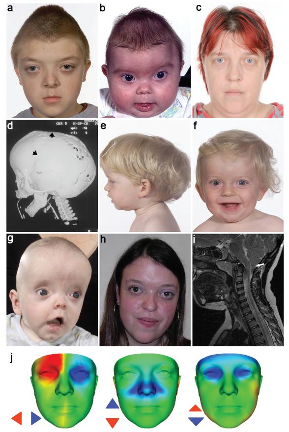
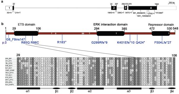
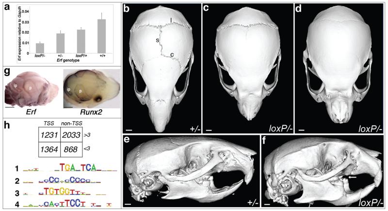
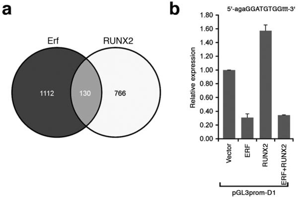
The extracellular signal-related kinases 1 and 2 (ERK1/2) are key proteins mediating mitogen-activated protein kinase signaling downstream of RAS: phosphorylation of ERK1/2 leads to nuclear uptake and modulation of multiple targets. Here, we show that reduced dosage of ERF, which encodes an inhibitory ETS transcription factor directly bound by ERK1/2 (refs. 2,3,4,5,6,7), causes complex craniosynostosis (premature fusion of the cranial sutures) in humans and mice. Features of this newly recognized clinical disorder include multiple-suture synostosis, craniofacial dysmorphism, Chiari malformation and language delay. Mice with functional Erf levels reduced to ∼30% of normal exhibit postnatal multiple-suture synostosis; by contrast, embryonic calvarial development appears mildly delayed. Using chromatin immunoprecipitation in mouse embryonic fibroblasts and high-throughput sequencing, we find that ERF binds preferentially to elements away from promoters that contain RUNX or AP-1 motifs. This work identifies ERF as a novel regulator of osteogenic stimulation by RAS-ERK signaling, potentially by competing with activating ETS factors in multifactor transcriptional complexes.


Figures

Figure 1
Figure 1
Clinical features of subjects heterozygous for ERF mutations. (a-d) Family 1 showing subject IV-1 aged 10 yr, in whom exome sequencing was performed (a), his brother IV-2 aged 4 mo (b) and mother III-3 aged 37 yr (c). The computed tomographic head scan of IV-2 aged 5 mo (d) shows synostosis of the left coronal and sagittal sutures (arrowheads) associated with multiple craniolacunae; the lambdoid and squamosal sutures remain patent. (e-h) Subjects identified in follow-up sequencing had clinical diagnoses ranging from non-syndromic sagittal (e, III-1 in Family 5 aged 1.6 yr) or unilateral lambdoid synostosis (f, III-1 in Family 4 aged 1.2 yr) to FGFR2 mutation-negative Crouzon syndrome (g, II-1 in Family 10 aged 4 mo; h, III-1 in Family 7 aged 18 yr). (i) Magnetic resonance brain imaging (sagittal, T1 view) of III-1 in Family 4 aged 7.1 yr, showing Chiari malformation (12 mm herniation of cerebellar tonsils through the foramen magnum, arrow). (j) Comparison of average faces between ERF-mutant (n = 14) and control (n = 381) subjects. Red/blue denotes normalized displacement at over 1.5 SD, highlighting shared features of hypertelorism (left), vertical nasal displacement (centre) and prominent forehead with exorbitism (right). Written consent was provided for the publication of all photographs.
Figure 2
Figure 2
Exon and domain structure of ERF and mutations identified in craniosynostosis. ERF comprizes 4 exons (a) extending over 7.6 kb and encodes a 548 amino acid protein (b). The positions of serine (S) and threonine (T) sites phosphorylated by ERK are indicated. Two missense substitutions p.Arg65Gln and p.Arg86Cys localize to the ETS DNA-binding domain. The lineup in the bottom panel shows the ETS domain sequence in a representative member of each ETS subfamily from humans. Fully and partially conserved residues are filled dark and light grey, respectively.
Figure 3
Figure 3
Analysis of Erf in mouse mutants and embryonic fibroblasts. (a) Quantitative RT-PCR of Erf in E16.5 calvariae of different genotypes, showing reduced expression of ErfloxP relative to wild type Erf allele. Error bars indicate standard error of mean. (b-f), Micro-CT scanning of heads of mice aged 9 weeks. Note normal morphology and patent sagittal (s), coronal (c) and lambdoid (l) sutures in the Erf+/− mutant (b,e), whereas the ErfloxP/− littermates have craniosynostosis of the sagittal and coronal sutures (c) or sagittal, coronal and lambdoid sutures (d). Note dental malocclusion (arrow) on the side view (f) of skull shown in d. Scale bars: 1.12 mm (b,e), 1.01 mm (c,d,f). (g) Whole mount RNA in situ hybridization of Erf (left) and Runx2 (right) in wild type E16.5 mouse calvariae. Note similar expression patterns coinciding with osteogenic fronts of parietal (p) and interparietal (ip) bones. Scale bars: 1 mm. (h) Summary of ChIP-Seq analysis using antibody to Erf in mouse embryonic fibroblasts. In the upper panel, box shows number of peaks identified according to whether they were located within 1 kb of a transcription start site (TSS) and whether they showed loss of binding in the presence of FCS (−FCS/+FCS >3). In the lower panel, MEME analysis of the 2033 non-TSS dynamically bound peaks (>3) identifies enrichment for motifs corresponding to binding sites for AP-1 (#1: 5′-TGANTCA-3′), RUNX (#3: 5′-TGTGG-3′) and ETS (#4: 5′-TTCCT-3′). Motif #2 was also observed in TSS peaks (Supplementary Fig. 9a).
Figure 4
Figure 4
Overlapping transcriptional targets of Erf and RUNX2 identified by ChIP-Seq. (a) Comparison of −FCS/+FCS >3, non-TSS murine Erf targets identified in this work (n = 2033) with orthologous human RUNX2 targets identified by Little et al. (n = 1603). To improve the specificity of linkage to regulated genes, ChIP-Seq peaks more than 40 kb from the closest RefSeq gene were excluded. Peaks were assigned to the gene with the closest exon. (b) Transactivation analysis using the hybrid Erf-Runx2 binding target identified by ChIP-Seq (sequence at top with core target capitalized). HeLa cells were transfected with 1 μg plasmid containing empty vector, ERF or RUNX2 cDNAs, or combinations as indicated. Results were normalized relative to empty vector and expressed as the mean ± SEM of 4 independent experiments.

References

    1. Plotnikov A, Zehorai E, Procaccia S, Seger R. The MAPK cascades: signaling components, nuclear roles and mechanisms of nuclear translocation. Biochim. Biophys. Acta. 2011;1813:1619–1633. - PubMed
    1. Mavrothalassitis GJ, Papas TS. Positive and negative factors regulate the transcription of the ETS2 gene via an oncogene-responsive-like unit within the ETS2 promoter region. Cell Growth Differ. 1991;2:215–224. - PubMed
    1. Sgouras DN, et al. ERF: an ETS domain protein with strong transcriptional repressor activity, can suppress ets-associated tumorigenesis and is regulated by phosphorylation during cell cycle and mitogenic stimulation. EMBO J. 1995;14:4781–4793. - PMC - PubMed
    1. Le Gallic L, Sgouras D, Beal G, Mavrothalassitis G. Transcriptional repressor ERF is a Ras/mitogen-activated protein kinase target that regulates cellular proliferation. Mol. Cell. Biol. 1999;19:4121–4133. - PMC - PubMed
    1. Le Gallic L, Virgilio L, Cohen P, Biteau B, Mavrothalassitis G. ERF nuclear shuttling, a continuous monitor of Erk activity that links it to cell cycle progression. Mol. Cell. Biol. 2004;24:1206–1218. - PMC - PubMed

Publication types

MeSH terms