. 2014 Feb;472(2):471-81.
doi: 10.1007/s11999-013-2788-y.

MRI predicts ALVAL and tissue damage in metal-on-metal hip arthroplasty

Affiliations

MRI predicts ALVAL and tissue damage in metal-on-metal hip arthroplasty

Danyal H Nawabi et al. Clin Orthop Relat Res. 2014 Feb.

Abstract

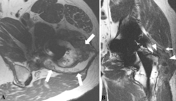
Background: Adverse local tissue reactions (ALTR) around metal-on-metal (MOM) hip arthroplasties are increasingly being recognized as a cause of failure. These reactions may be associated with intraoperative tissue damage and complication rates as high as 50% after revision. Although MRI can identify ALTR in MOM hips, it is unclear whether the MRI findings predict those at revision surgery.

Questions/purposes: We therefore (1) identified which MRI characteristics correlated with histologically confirmed ALTR (using the aseptic lymphocytic vasculitis-associated lesions [ALVAL] score) and intraoperative tissue damage and (2) developed a predictive model using modified MRI to detect ALVAL and quantify intraoperative tissue damage.

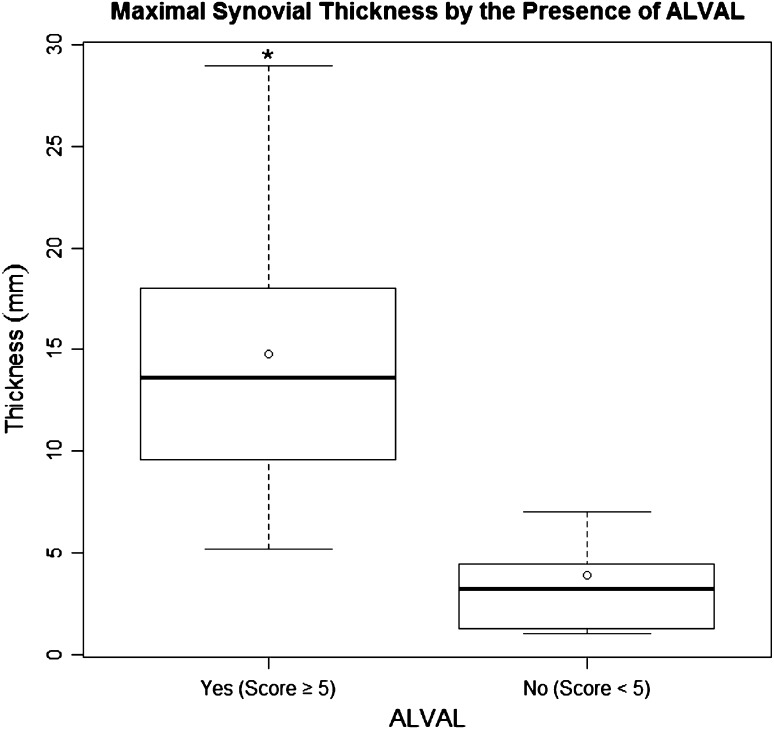
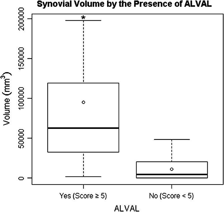
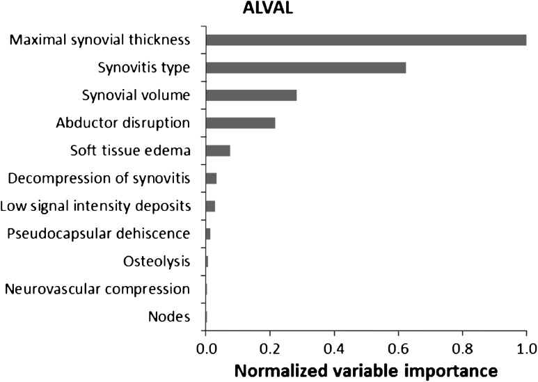
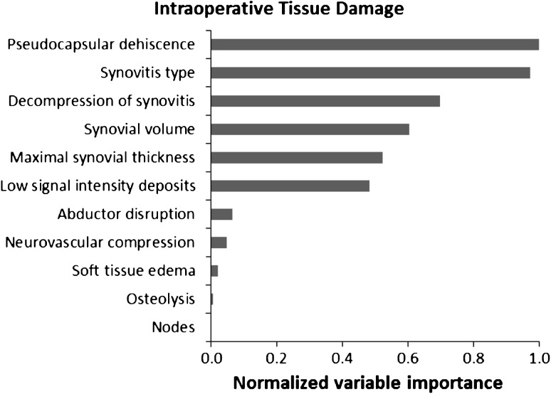
Methods: We retrospectively reviewed 68 patients with failed MOM hip arthroplasties who underwent preoperative MRI and subsequent revision surgery. Images were analyzed to determine synovial volume, osteolysis, and synovial thickness. The ALVAL score was used to grade tissue samples, thus identifying a subset of patients with ALTR. Intraoperative tissue damage was graded using a four-point scale. Random forest analysis determined the sensitivity and specificity of MRI characteristics in detecting ALVAL (score ≥ 5) and intraoperative tissue damage.

Results: Maximal synovial thicknesses and synovial volumes as determined on MRI correlated with the ALVAL score and were higher in cases of severe intraoperative tissue damage. Our MRI predictive model showed sensitivity and specificity of 94% and 87%, respectively, for detecting ALVAL and 90% and 86%, respectively, for quantifying intraoperative tissue damage.

Conclusions: MRI is sensitive and specific in detecting ALVAL and tissue damage in patients with MOM hip implants. MRI can be used as a screening tool to guide surgeons toward timely revision surgery.


Figures

Fig. 1A–B
Fig. 1A–B
(A) The FSE pulse sequence for a 29-year-old man after a right hip resurfacing arthroplasty shows marked susceptibility artifact (white arrowheads) arising from the cobalt-chromium implant, precluding observation of the synovial lining. (B) A coronal MAVRIC sequence shows marked reduction in susceptibility artifact, unmasking right hip synovitis (white arrows).
Fig. 2A–C
Fig. 2A–C
(A) An axial FSE image for a 51-year-old woman after right hip resurfacing arthroplasty shows mixed- (fluid and solid) type synovitis (white arrow). (B) The coronal MAVRIC prototype sequence for a 57-year-old man after a MOM THA shows marked synovial thickening (white arrow). (C) The axial FSE image for a 65-year-old man after his THA shows decompression of synovitis into the greater trochanteric bursa (white arrowhead). There is lateral dehiscence of the posterior pseudocapsule (white arrow). The overlaid graphics show the segmentation method used to quantify the volume of synovitis.
Fig. 3
Fig. 3
A coronal MAVRIC prototype pulse sequence for a 52-year-old woman obtained after right hip resurfacing arthroplasty shows extensive replacement of the normal periacetabular marrow fat by low signal intensity (white arrowheads), consistent with osteolysis. There is an adjacent large extracapsular low signal intensity deposit (white arrow) abutting the sciatic nerve.
Fig. 4
Fig. 4
A coronal FSE image obtained after a left MOM THA in a 62-year-old man shows decompression of the synovitis into the greater trochanteric bursa with associated disruption of the hip abductors and retraction of the torn tendons (white arrow). Atrophy of the muscle bellies also is evident (black arrow).
Fig. 5A–B
Fig. 5A–B
(A) An axial FSE image obtained after a right hip resurfacing arthroplasty in a 51-year-old woman shows marked anterior synovial expansion (white arrow) impinging the femoral nerve (white arrowhead). (B) The coronal FSE image for a 57-year-old man obtained after a right THA shows marked posterior synovial expansion (white arrow) abutting the adjacent sciatic nerve (white arrowheads).
Fig. 6
Fig. 6
A box plot shows the interquartile range (box edges representing 25th and 75th percentiles), median (dark bar inside box), and mean (circle) for synovial thickness by the presence of ALVAL. The error bars represent the minimum and maximum observed values within 1.5 times the interquartile range values. Outliers are not shown. The asterisk indicates a major difference in medians between outcome groups.
Fig. 7
Fig. 7
A box plot shows the interquartile range (box edges representing 25th and 75th percentiles), median (band inside box), and mean (circle) for synovial volume by the presence of ALVAL. The error bars represent the minimum and maximum observed values within 1.5 times the interquartile range values. Outliers are not shown. The asterisk indicates a major difference in medians between outcome groups.
Fig. 8A–B
Fig. 8A–B
(A) An axial (A) FSE image obtained after MOM THA in a 57-year-old man shows marked synovial expansion and thickening (white arrows) with anterior and posterior decompression. (B) The patient’s coronal FSE image shows a large volume of synovitis decompressing inferiorly (white arrows) and superiorly into the subiliacus bursa (black arrowheads). The high volume of mixed- (fluid and solid) type synovitis combined with the greatly thickened synovial lining and disruption of the abductors resulting from distention of the pseudocapsule is suggestive of a high ALVAL score. This was confirmed at revision surgery with subsequent histologic analysis showing an ALVAL score of 9.
Fig. 9A–B
Fig. 9A–B
(A) The axial FSE image obtained after a left MOM THA in a 46-year-old man shows synovial expansion and thickening with decompression of synovitis into the greater trochanteric bursa (white arrows) through a lateral dehiscence in the pseudocapsule. (B) The patient’s coronal FSE image shows synovial expansion (white arrowheads) undermining the hip abductors with resulting disruption of the anterolateral fibers of the gluteus medius (small white arrow). The mixed-type synovial expansion with associated pseudocapsular dehiscence and extensive decompression of synovitis suggests substantial intraoperative damage. This is further supported by the large volume of synovitis and thick synovial lining. These findings were confirmed at revision surgery, when substantial soft tissue damage was seen and the patient received an intraoperative damage score of 3.
Fig. 10
Fig. 10
A random forest analysis shows the normalized importance of MRI characteristics in predicting the presence of ALVAL. Importance is normalized to the best predictor, assigned the value of 1.
Fig. 11
Fig. 11
A random forest analysis shows the normalized importance of MRI characteristics in predicting the presence of intraoperative tissue damage. Importance is normalized to the best predictor, assigned the value of 1.

References

    1. Amstutz HC, Le Duff MJ, Campbell PA, Wisk LE, Takamura KM. Complications after metal-on-metal hip resurfacing arthroplasty. Orthop Clin North Am. 2011;42:207–230. doi: 10.1016/j.ocl.2010.12.002. - DOI - PubMed
    1. Campbell P, Ebramzadeh E, Nelson S, Takamura K, De Smet K, Amstutz HC. Histological features of pseudotumor-like tissues from metal-on-metal hips. Clin Orthop Relat Res. 2010;468:2321–2327. doi: 10.1007/s11999-010-1372-y. - DOI - PMC - PubMed
    1. Coulter G, Young DA, Dalziel RE, Shimmin AJ. Birmingham hip resurfacing at a mean of ten years: results from an independent centre. J Bone Joint Surg Br. 2012;94:315–321. doi: 10.1302/0301-620X.94B3.28185. - DOI - PubMed
    1. Dorr LD, Wan Z, Longjohn DB, Dubois B, Murken R. Total hip arthroplasty with use of the Metasul metal-on-metal articulation: four to seven-year results. J Bone Joint Surg Am. 2000;82:789–798. - PubMed
    1. Glyn-Jones S, Pandit H, Kwon YM, Doll H, Gill HS, Murray DW. Risk factors for inflammatory pseudotumour formation following hip resurfacing. J Bone Joint Surg Br. 2009;91:1566–1574. doi: 10.1302/0301-620X.91B12.22287. - DOI - PubMed

MeSH terms

Supplementary concepts