Review
. 2013 Jun;8(3):494-509.
doi: 10.1007/s11481-013-9434-z. Epub 2013 Jan 27.

Microglia: key elements in neural development, plasticity, and pathology

Affiliations
Review

Microglia: key elements in neural development, plasticity, and pathology

Ukpong B Eyo et al. J Neuroimmune Pharmacol. 2013 Jun.

Abstract

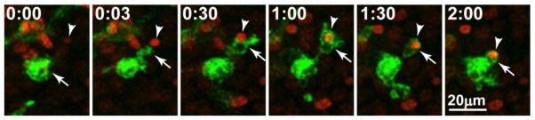
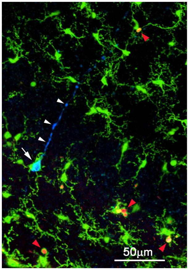
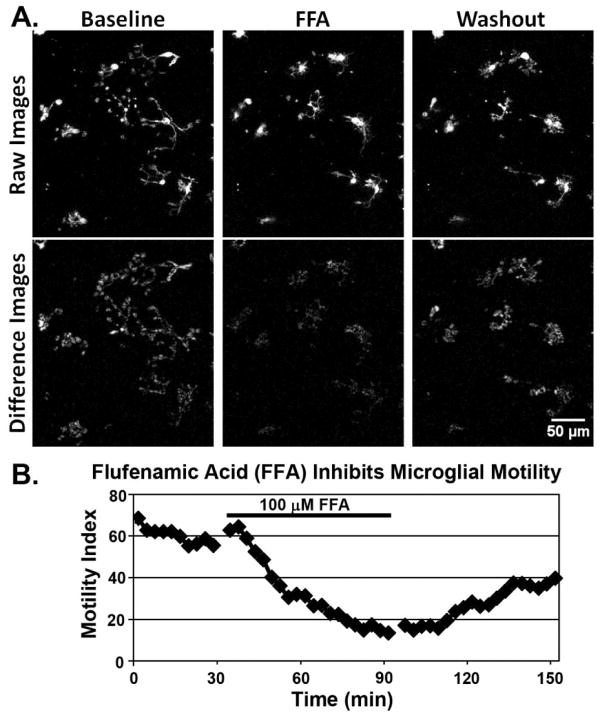
A century after Cajal identified a "third element" of the nervous system, many issues have been clarified about the identity and function of one of its major components, the microglia. Here, we review recent findings by microgliologists, highlighting results from imaging studies that are helping provide new views of microglial behavior and function. In vivo imaging in the intact adult rodent CNS has revolutionized our understanding of microglial behaviors in situ and has raised speculation about their function in the uninjured adult brain. Imaging studies in ex vivo mammalian tissue preparations and in intact model organisms including zebrafish are providing insights into microglial behaviors during brain development. These data suggest that microglia play important developmental roles in synapse remodeling, developmental apoptosis, phagocytic clearance, and angiogenesis. Because microglia also contribute to pathology, including neurodevelopmental and neurobehavioral disorders, ischemic injury, and neuropathic pain, promising new results raise the possibility of leveraging microglia for therapeutic roles. Finally, exciting recent work is addressing unanswered questions regarding the nature of microglial-neuronal communication. While it is now apparent that microglia play diverse roles in neural development, behavior, and pathology, future research using neuroimaging techniques will be essential to more fully exploit these intriguing cellular targets for effective therapeutic intervention applied to a variety of conditions.


Conflict of interest statement

The authors declare that they have no conflict of interest.

Figures

Figure 1
Figure 1. Morphology of ‘resting/surveillant’ microglia in developing mouse neocortex
Confocal image shows GFP-expressing microglia (green) and YFP-expressing pyramidal neurons (yellow) in cortical tissue from an early postnatal double-transgenic reporter mouse (CX3CR1GFP/+:Thy1-YFP). Note the fine microglia branches intercalated among the dendritic arbors of the pyramidal neurons.
Figure 2
Figure 2. Microglial Ramification from Development to Adulthood
A, Microglia from a neonatal mouse hippocampus showing a central soma and several projections. B, Schematic representation of the progressive ramification of microglia through development into adulthood. Brain-resident microglia are first observed during embryonic development as amoeboid cells with few projections. Beginning prenatally and extending into the early postnatal period, microglia begin to transform into cells with more defined primary projections with little secondary branching. By the second postnatal week onwards, microglial morphology consists of more elaborate primary, secondary and even tertiary branching in their projections.
Figure 3
Figure 3. Microglial motility facilitates rapid mobilization to injured neurons
In this case, injury was experimentally induced in a mouse hippocampal tissue slice by focal laser burn. Within minutes after the injury, an activated amoeboid microglial cell (arrow) extends a branch toward a Sytox-labeled nucleus of an injured neuron (arrowhead), contacts it, and engulfs it. Time is shown in hr:min.
Figure 4
Figure 4. Microglial engagement of early and late stage apoptotic cells in the developing mouse hippocampus
Three-channel confocal image showing primitive ramified microglia (green), cleaved caspase-3 immunostained early apoptotic pyramidal neuron (blue), and PSVue-stained late stage apoptotic bodies (red). A microglial cell (white arrow) enwraps the soma of an apoptotic pyramidal neuron labeled by CC3 antibodies. Note labeling of the primary apical dendrite (white arrowheads), which has a blebby, degenerating appearance. Other microglia nearby are in the process of phagocytosing late stage apoptotic bodies labeled with PSVue+ (red arrowheads). The cells are dying by natural, developmental apoptosis.
Figure 5
Figure 5. Pharmacological modulation of microglial motility by a non-steroidal anti-inflammatory drug (NSAID), flufenamic acid (FFA)
(A) Time-lapse imaging of microglia in a live brain tissue slice from a GFP-reporter mouse shows that FFA (100 μM) reversibly inhibits microglial motility. Top row shows raw fluorescence images. Bottom row shows ‘difference images,’ which indicate motile changes between sequential time-points in an image sequence. Note the lack of motility during FFA. (B) Quantification of the motility index (MI), based on the difference images, shows slow decline during FFA and recovery during washout. For details of methods, see Eyo & Dailey (2012). FFA has been shown to block extension of processes from microglia (Davalos et al., 2005; Wu et al., 2007) and astrocytes (Kim and Dustin, 2006) after laser injury. FFA may inhibit microglial motility by blocking connexin hemichannels. FFA is an anthranilic acid derivative with analgesic, anti-inflammatory, and antipyretic properties.
Figure 6
Figure 6. Time-lapse multiphoton imaging sequences showing rapid mobilization of microglia to injured neurons in a live brain tissue slice from a neonatal mouse lacking the P2X7 receptor (P2X7−/−)
Microglia cell bodies and branches (green) are visible due to expression of green fluorescent protein (GFP) in this mouse line (P2X7−/−:CX3CR1GFP/+). The many healthy cells in the tissue are unlabeled. (A) Time-lapse movie under baseline conditions (00:00–00:17) show continual remodeling of microglial branches. Focal tissue injury (00:17) was induced along a line by brief exposure to high intensity laser light (white line between white arrowheads). Within minutes, injured cells begin to take up a membrane-impermeable red fluorescent DNA-binding dye, ToPro3, and nearby microglia extend branches (yellow arrowheads) toward the laser-damaged cells (00:28). Within a couple of hours, stationary microglia have transformed to activated fusiform or amoeboid microglia that migrate and accumulate near the injured cells (01:47). (B) Higher-magnification view of the same time-lapse sequence shows migration of a microglia cell (yellow arrowheads) toward the injured cells. Microglia respond to tissue injury even though they lack the P2X7 purinoceptor in these knockout mice. Time is shown in hr:min. See supplementary Movie 4.

References

    1. Araque A, Parpura V, Sanzgiri RP, Haydon PG. Tripartite synapses: glia, the unacknowledged partner. Trends Neurosci. 1999;22:208–215. - PubMed
    1. Ashwell KW, Hollander H, Streit W, Stone J. The appearance and distribution of microglia in the developing retina of the rat. Vis Neurosci. 1989;2:437–448. - PubMed
    1. Barreto G, White RE, Ouyang Y, Xu L, Giffard RG. Astrocytes: targets for neuroprotection in stroke. Cent Nerv Syst Agents Med Chem. 2011;11:164–173. - PMC - PubMed
    1. Battisti WP, Wang J, Bozek K, Murray M. Macrophages, microglia, and astrocytes are rapidly activated after crush injury of the goldfish optic nerve: a light and electron microscopic analysis. J Comp Neurol. 1995;354:306–320. - PubMed
    1. Beers DR, Henkel JS, Xiao Q, Zhao W, Wang J, Yen AA, Siklos L, McKercher SR, Appel SH. Wild-type microglia extend survival in PU.1 knockout mice with familial amyotrophic lateral sclerosis. Proc Natl Acad Sci U S A. 2006;103:16021–16026. - PMC - PubMed

Publication types

LinkOut - more resources