

Increased generation of cyclopentenone prostaglandins after brain ischemia and their role in aggregation of ubiquitinated proteins in neurons
- PMID: 23355003
- PMCID: PMC3692569
- DOI: 10.1007/s12640-013-9377-4
Increased generation of cyclopentenone prostaglandins after brain ischemia and their role in aggregation of ubiquitinated proteins in neurons
Abstract
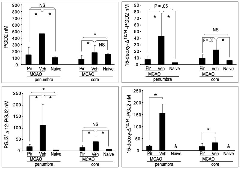
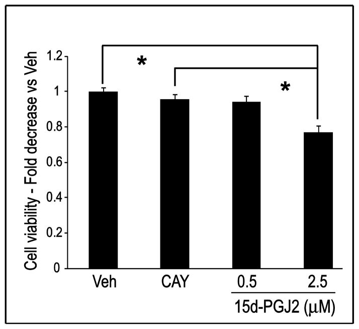
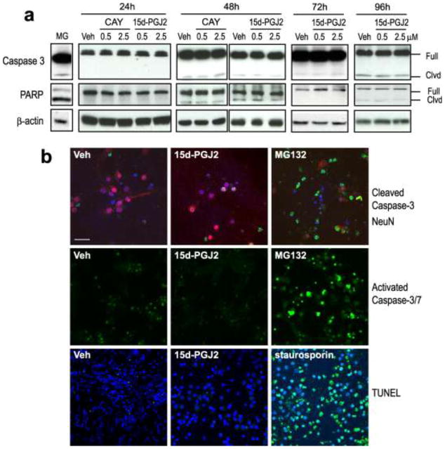
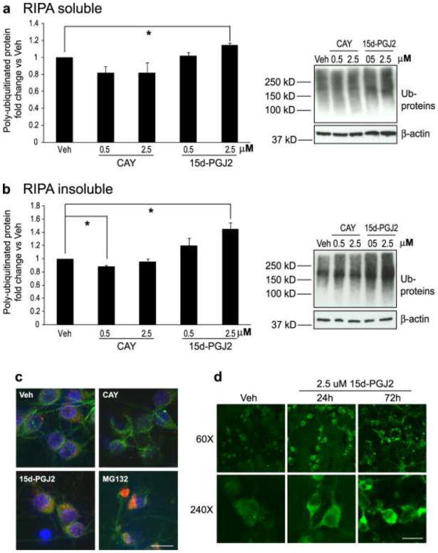
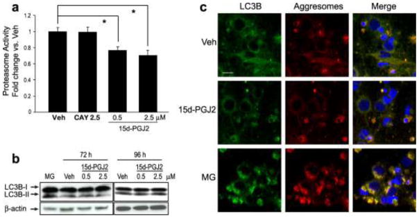
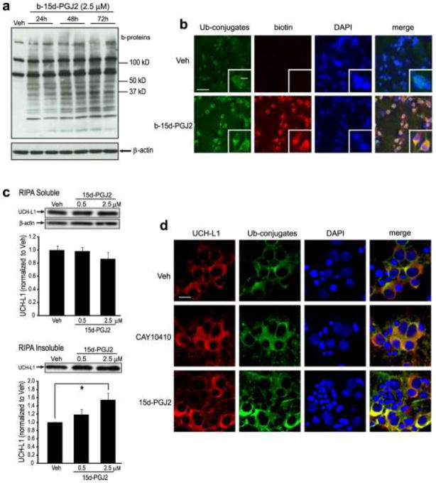
The cyclopentenone prostaglandin (CyPG) J₂ series, including prostaglandin J₂ (PGJ₂), Δ¹²-PGJ₂, and 15-deoxy-∆¹²,¹⁴-prostaglandin J₂ (15d-PGJ₂), are active metabolites of PGD₂, exerting multiple effects on neuronal function. However, the physiologic relevance of these effects remains uncertain as brain concentrations of CyPGs have not been precisely determined. In this study, we found that free PGD₂ and the J₂ series CyPGs (PGJ₂, Δ¹²-PGJ₂, and 15d-PGJ₂) were increased in post-ischemic rat brain as detected by UPLC-MS/MS with 15d-PGJ₂ being the most abundant CyPG. These increases were attenuated by pre-treating with the cyclooxygenase (COX) inhibitor piroxicam. Next, effects of chronic exposure to 15d-PGJ₂ were examined by treating primary neurons with 15d-PGJ₂, CAY10410 (a 15d-PGJ₂ analog lacking the cyclopentenone ring structure), or vehicle for 24 to 96 h. Because we found that the concentration of free 15d-PGJ₂ decreased rapidly in cell culture medium, freshly prepared medium containing 15d-PGJ₂, CAY10410, or vehicle was changed twice daily to maintain steady extracellular concentrations. Incubation with 2.5 μM 15d-PGJ₂, but not CAY10410, increased the neuronal cell death without the induction of caspase-3 or PARP cleavage, consistent with a primarily necrotic mechanism for 15d-PGJ₂-induced cell death which was further supported by TUNEL assay results. Ubiquitinated protein accumulation and aggregation was observed after 96 h 15d-PGJ₂ incubation, accompanied by compromised 20S proteasome activity. Unlike another proteasome inhibitor, MG132, 15d-PGJ₂ treatment did not activate autophagy or induce aggresome formation. Therefore, the cumulative cytotoxic effects of increased generation of CyPGs after stroke may contribute to delayed post-ischemic neuronal injury.
Conflict of interest statement
The authors declare no conflict of interest. The contents do not represent the views of the Department of Veterans Affairs or the United States Government.
Figures
Similar articles
-
The biphasic effects of cyclopentenone prostaglandins, prostaglandin J(2) and 15-deoxy-Delta(12,14)-prostaglandin J(2) on proliferation and apoptosis in rat basophilic leukemia (RBL-2H3) cells.Biochem Pharmacol. 2004 Apr 1;67(7):1259-67. doi: 10.1016/j.bcp.2003.10.037. Biochem Pharmacol. 2004. PMID: 15013841
-
An endogenous electrophile that modulates the regulatory mechanism of protein turnover: inhibitory effects of 15-deoxy-Delta 12,14-prostaglandin J2 on proteasome.Biochemistry. 2003 Dec 2;42(47):13960-8. doi: 10.1021/bi035215a. Biochemistry. 2003. PMID: 14636064
-
Effects of 15-deoxy-delta 12, 14-prostaglandin J2 on the expression of p53 in MCF-7 cells.Ann N Y Acad Sci. 2009 Aug;1171:202-9. doi: 10.1111/j.1749-6632.2009.04913.x. Ann N Y Acad Sci. 2009. PMID: 19723057
-
Peroxisome proliferator-activated receptor gamma (PPARgamma) ligands as bifunctional regulators of cell proliferation.Biochem Pharmacol. 2003 Oct 15;66(8):1381-91. doi: 10.1016/s0006-2952(03)00488-x. Biochem Pharmacol. 2003. PMID: 14555212 Review.
-
Cyclopentenone prostaglandins: new insights on biological activities and cellular targets.Med Res Rev. 2001 May;21(3):185-210. doi: 10.1002/med.1006. Med Res Rev. 2001. PMID: 11301410 Review.
Cited by
-
Role of UCHL1 in the pathogenesis of neurodegenerative diseases and brain injury.Ageing Res Rev. 2023 Apr;86:101856. doi: 10.1016/j.arr.2023.101856. Epub 2023 Jan 19. Ageing Res Rev. 2023. PMID: 36681249 Free PMC article. Review.
-
The Role of Ubiquitin-Proteasome Pathway and Autophagy-Lysosome Pathway in Cerebral Ischemia.Oxid Med Cell Longev. 2020 Jan 30;2020:5457049. doi: 10.1155/2020/5457049. eCollection 2020. Oxid Med Cell Longev. 2020. PMID: 32089771 Free PMC article. Review.
-
Thorough overview of ubiquitin C-terminal hydrolase-L1 and glial fibrillary acidic protein as tandem biomarkers recently cleared by US Food and Drug Administration for the evaluation of intracranial injuries among patients with traumatic brain injury.Acute Med Surg. 2021 Jan 19;8(1):e622. doi: 10.1002/ams2.622. eCollection 2021 Jan-Dec. Acute Med Surg. 2021. PMID: 33510896 Free PMC article. Review.
-
L-PGDS-PGD2-DP1 Axis Regulates Phagocytosis by CD36+ MGs/MΦs That Are Exclusively Present Within Ischemic Areas After Stroke.Cells. 2024 Oct 20;13(20):1737. doi: 10.3390/cells13201737. Cells. 2024. PMID: 39451255 Free PMC article.
-
PACAP27 prevents Parkinson-like neuronal loss and motor deficits but not microglia activation induced by prostaglandin J2.Biochim Biophys Acta. 2014 Sep;1842(9):1707-19. doi: 10.1016/j.bbadis.2014.06.020. Epub 2014 Jun 23. Biochim Biophys Acta. 2014. PMID: 24970746 Free PMC article.
References
-
- Andersson FI, Werrell EF, McMorran L, Crone WJ, Das C, Hsu ST, Jackson SE. The effect of Parkinson’s-disease-associated mutations on the deubiquitinating enzyme UCH-L1. Journal of molecular biology. 2011;407:261–272. - PubMed
Publication types
MeSH terms
Substances
Grants and funding
LinkOut - more resources
Full Text Sources
Other Literature Sources
Research Materials
