

Innovative endovascular technique for treatment of rare cause of haemoptysis in young
- PMID: 23355588
- PMCID: PMC3604488
- DOI: 10.1136/bcr-2012-008205
Innovative endovascular technique for treatment of rare cause of haemoptysis in young
Abstract
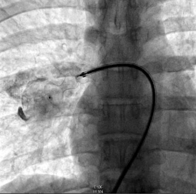
A 17-year-old boy reported for evaluation of two episodes of massive haemoptysis. His clinical examination was unremarkable and investigations (haemogram, coagulogram, serological tests for connective tissue disorders) were normal. A 64-slice CT angiography revealed a saccular aneurysm of 3.8×3.7×3.3 cm arising from the right lower lobe pulmonary artery which was confirmed by cardiac catheterisation. The aneurysm was successfully blocked with a 16-14 Amplatzer duct occluder. A repeat CT angiogram performed after 15 days revealed the device in situ obliterating the aneurysm. Pulmonary artery aneurysm is an extremely rare cause of massive haemoptysis and indicates imminent rupture of the aneurysm which can be rapidly fatal. This case highlights the importance of using an innovative endovascular technique for treatment of a rare cause of haemoptysis.
Figures


Similar articles
-
Haemoptysis in pulmonary artery aneurysm associated with pulmonary hypertension: surgical dilemma.Aust N Z J Surg. 1982 Dec;52(6):557-9. doi: 10.1111/j.1445-2197.1982.tb06110.x. Aust N Z J Surg. 1982. PMID: 6962723 No abstract available.
-
Subclavian artery aneurysm: a rare cause of massive haemoptysis.BMJ Case Rep. 2021 Mar 9;14(3):e241225. doi: 10.1136/bcr-2020-241225. BMJ Case Rep. 2021. PMID: 33687930 Free PMC article.
-
Saccular aneurysm within a persistent ductus arteriosus.Lancet. 2012 Feb 18;379(9816):e33. doi: 10.1016/S0140-6736(11)61352-4. Epub 2011 Dec 15. Lancet. 2012. PMID: 22177260 No abstract available.
-
Saphenous vein graft aneurysm - Unusual cause of hemoptysis: A case report and review of literature.J Postgrad Med. 2020 Jul-Sep;66(3):165-168. doi: 10.4103/jpgm.JPGM_187_20. J Postgrad Med. 2020. PMID: 32675454 Free PMC article. Review.
-
[An operative case of bilateral peripheral pulmonary arterial aneurysms].Kyobu Geka. 1997 Apr;50(4):331-4. Kyobu Geka. 1997. PMID: 9095597 Review. Japanese.
Cited by
-
Mediastinal bronchial artery aneurysm with short inflow segment successfully treated with a patent ductus arteriosus occluder device.J Vasc Surg Cases Innov Tech. 2020 Feb 20;6(1):93-95. doi: 10.1016/j.jvscit.2019.12.005. eCollection 2020 Mar. J Vasc Surg Cases Innov Tech. 2020. PMID: 32095664 Free PMC article.
References
-
- Lopez-Candales A, Kleiger RE, Aleman-Gomez J, et al. Pulmonary artery aneurysm: review and case report. Clin Cardiol 1995;18:738–40 - PubMed
-
- Karkoulias K, Lykouras D, Nanopoulou M, et al. An unexpected pulmonary arterial aneurysm in a COPD patient. Acta Clin Belg 2011;66:379–80 - PubMed
-
- Lee R, Son JS, Park YM. A case of left main pulmonary artery aneurysm associated with valvular pulmonary stenosis in a child. Pediatr Cardiol 2011;32:1055–6 - PubMed
-
- Goda M, Budts W, Troost E, et al. Bicuspid pulmonary valve with atrial septal defect leading to pulmonary aneurysm. Ann Thorac Surg 2012;93:1706–8 - PubMed
-
- Vistarini N, Aubert S, Gandjbakhch I, et al. Surgical treatment of a pulmonary artery aneurysm. Eur J Cardiothorac Surg 2007;31:1139–41 - PubMed
Publication types
MeSH terms
LinkOut - more resources
Full Text Sources
Other Literature Sources
Medical