. 2013 Apr;41(7):e77.
doi: 10.1093/nar/gkt002. Epub 2013 Jan 25.

A red/far-red light-responsive bi-stable toggle switch to control gene expression in mammalian cells

Affiliations

A red/far-red light-responsive bi-stable toggle switch to control gene expression in mammalian cells

Konrad Müller et al. Nucleic Acids Res. 2013 Apr.

Abstract

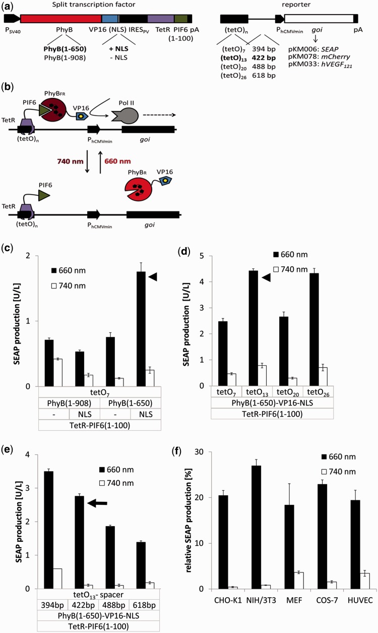
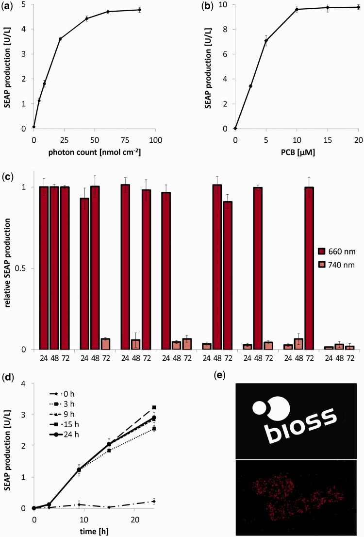
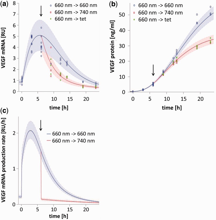
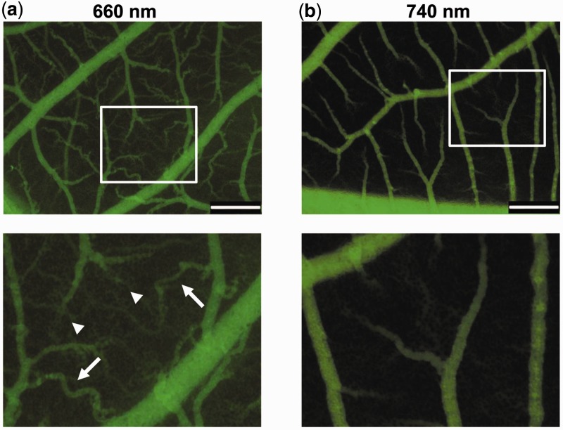
Growth and differentiation of multicellular systems is orchestrated by spatially restricted gene expression programs in specialized subpopulations. The targeted manipulation of such processes by synthetic tools with high-spatiotemporal resolution could, therefore, enable a deepened understanding of developmental processes and open new opportunities in tissue engineering. Here, we describe the first red/far-red light-triggered gene switch for mammalian cells for achieving gene expression control in time and space. We show that the system can reversibly be toggled between stable on- and off-states using short light pulses at 660 or 740 nm. Red light-induced gene expression was shown to correlate with the applied photon number and was compatible with different mammalian cell lines, including human primary cells. The light-induced expression kinetics were quantitatively analyzed by a mathematical model. We apply the system for the spatially controlled engineering of angiogenesis in chicken embryos. The system's performance combined with cell- and tissue-compatible regulating red light will enable unprecedented spatiotemporally controlled molecular interventions in mammalian cells, tissues and organisms.


Figures

Figure 1.
Figure 1.
Design, optimization and validation of the red light-inducible transgene expression system. (a) Configuration of the red light-inducible expression system. The building blocks for the split transcription factor are encoded on a bicistronic expression vector under the control of the simian virus 40 promoter PSV40. In the first cistron, the N-terminal fragments of PhyB (amino acids 1–650 or 1–908) are fused to VP16 and optionally to an NLS. In the second cistron, the N-terminal 100 amino acids of PIF6 are fused to the tetracycline repressor TetR. Translation of the second cistron is induced by a polioviral internal ribosome entry site, IRESPV. The response vectors comprise multiple repeats of the TetR-specific operator tetO fused via spacers of different length to the minimal human cytomegalovirus immediate early promoter PhCMVmin. This chimeric promoter was configured to control expression of different genes of interest (goi): SEAP, secreted alkaline phosphatase; hVEGF121, 121 amino acid splice variant of human vascular endothelial growth factor or the fluorescent protein mCherry. (b) Mode of function. Red light illumination converts PhyB into the FR form (PhyBFR) and induces heterodimerization with PIF6 tethered via TetR to the tetOn operator site. The PhyB-fused VP16 domain recruits the transcription initiation complex and triggers activation of the minimal promoter PhCMVmin. Absorption of a far-red photon (740 nm) converts PhyB into the R form (PhyBR) and triggers dissociation from PIF6, thereby resulting in de-activation of the target promoter and transcriptional silence. (c–e) Optimization of the red light-inducible expression system in CHO-K1. The indicated configurations of the red light-inducible expression system were transfected into 70 000 CHO-K1 cells. After incubation for 24 h, medium containing 15 µM PCB was added. After 1 h incubation in the dark, the cells were exposed for 24 h to red (660 nm) or far-red (740 nm) light before quantification of the reporter gene SEAP. (c) Impact of PhyB variant (908 or 650 amino acids) and the absence or presence of an NLS. (d) Impact of the tetO copy number. (e) Impact of spacer length between the operator and the minimal promoter. Arrowheads mark the configurations that were optimized in the subsequent experiments. The configuration showing the best induction ratio (arrow) was used for the subsequent studies. (f) Characterization of red light-inducible gene expression in different mammalian cell lines. Plasmids pKM022 and pKM006 were transfected into CHO-K1, mouse fibroblasts (NIH/3T3, MEF), monkey fibroblasts (COS-7) and primary HUVEC. After 24 h, medium containing 15 µM PCB was added. The cells were subsequently incubated for 1 h in the dark, and further cultivated under 660 or 740 nm light for 24 h before quantification of SEAP production. To correct for different transfection efficiencies for the different cell lines, the expression data were normalized to SEAP expression levels under the control of the tetracycline-inducible expression system (vectors pSAM200 and pKM006, Table 1). Data are means of four independent experiments, and error bars indicate the standard deviation.
Figure 2.
Figure 2.
Detailed characterization of red light-inducible gene expression. (a–d) 70 000 CHO-K1 cells were transfected with the red light-inducible SEAP expression system (plasmids pKM006 and pKM022). After 24 h, the medium was replaced by fresh medium containing PCB (15 µM unless stated otherwise). The cells were cultivated for 1 h in the dark and subsequently subjected to the indicated illumination conditions before quantification of SEAP production. (a) Dose–response curve for red light-inducible SEAP expression. Cells were exposed to a 25 min pulse of 660 nm light at different intensities, resulting in the indicated photon number. After 24 h incubation in the dark, SEAP production was quantified. (b) PCB-adjustable transgene expression levels. Cells were exposed to a 25 min pulse of 660 nm light, resulting in a photon number of 80 nmol cm−2 for full induction of gene expression in the presence of increasing PCB concentrations. Subsequently, the cells were cultivated in the dark for 24 h before the quantification of SEAP production. (c) Reversibility of red light-inducible gene expression. Every 24 h, the cell culture medium was replaced by fresh PCB-containing medium, and the cells were illuminated at the indicated wavelengths. SEAP production was measured every 24 h. To correct for changes in gene expression over time because of cell growth, expression levels were normalized to the configuration, where cells were constantly kept under 660 nm light. (d) Effect of dark reversion of the PhyB–PIF6 interaction on transgene expression. Cells were grown under 660 nm light for the indicated periods before moving the cells to the dark. At the indicated time points, SEAP production was quantified. (e) Spatially resolved control of gene expression. CHO-K1 cells transgenic for red light-inducible mCherry expression (plasmids pKM022 and pKM078) were illuminated through a photo mask (top image) for 1 h (0.5 µmol m−2 s−1). After 23 h incubation in the dark, mCherry production was visualized. Scale bar, 1 cm. In Figure 2a–d, data represent the mean of four independent experiments, and error bars indicate the standard deviations.
Figure 3.
Figure 3.
Model-based analysis of the light-responsive gene expression kinetics. In all, 70 000 CHO-K1 cells were transfected for red-light inducible hVEGF121 expression (plasmids pKM022 and pKM033). After 24 h, the medium was supplemented with 15 µM PCB (t = −1 h). Cells were subsequently illuminated for 6 h at 660 nm and were then either switched to 660 nm, 740 nm or tetracycline-containing medium (arrow). (a) Switch-off kinetics of hVEGF121 mRNA. At the indicated points in time, cells were lysed, and hVEGF121 mRNA was quantified. (b) Switch-off kinetics of hVEGF121 protein production. At the indicated points in time, hVEGF121 was quantified in the cell culture supernatant. (c) Model prediction for the de novo synthesis of hVEGF121 mRNA. In (a) and (b), the shaded error bands are based on the error model detailed in Equation (9). The shaded bands in (c) indicate the 95% prediction confidence interval, which was calculated by propagating the uncertainties of the estimated parameters.
Figure 4.
Figure 4.
In vivo red light-controlled vascularization in chicken embryos. CHO-K1 cells were engineered for red light-inducible expression of the 121 amino acid splice variant of human vascular endothelial growth factor hVEGF121. One million cells were embedded in 3% polyethylene glycol gel and were applied onto the CAM of 9 days old chicken embryos. After illumination at (a) 660 or (b) 740 nm for 48 h, the microvasculature was visualized by injection of FITC–dextran and video imaging (see Supplementary Material for videos). Brush- and delta-like endpoint patterns (arrowheads) and tortuously shaped vascularization (arrows) are indicated. Top panels, overview; lower panels, insets at higher magnification. Scale bar, 500 µm.

Similar articles

Cited by

References

    1. Weber W, Fussenegger M. Emerging biomedical applications of synthetic biology. Nat. Rev. Genet. 2012;13:21–35. - PMC - PubMed
    1. Wang X, Chen X, Yang Y. Spatiotemporal control of gene expression by a light-switchable transgene system. Nat. Methods. 2012;9:266–269. - PubMed
    1. Yazawa M, Sadaghiani AM, Hsueh B, Dolmetsch RE. Induction of protein-protein interactions in live cells using light. Nat. Biotechnol. 2009;27:941–945. - PubMed
    1. Ye HF, Daoud-El Baba M, Peng RW, Fussenegger M. A synthetic optogenetic transcription device enhances blood-glucose homeostasis in mice. Science. 2011;332:1565–1568. - PubMed
    1. Khanna R, Huq E, Kikis EA, Al-Sady B, Lanzatella C, Quail PH. A novel molecular recognition motif necessary for targeting photoactivated phytochrome signaling to specific basic helix-loop-helix transcription factors. Plant Cell. 2004;16:3033–3044. - PMC - PubMed

Publication types

Substances