. 2013 Mar 1;41(5):3373-85.
doi: 10.1093/nar/gkt023. Epub 2013 Jan 25.

Molecular paleontology: a biochemical model of the ancestral ribosome

Affiliations

Molecular paleontology: a biochemical model of the ancestral ribosome

Chiaolong Hsiao et al. Nucleic Acids Res. .

Abstract

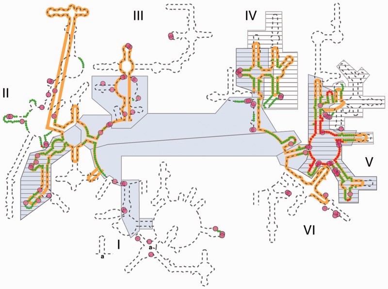
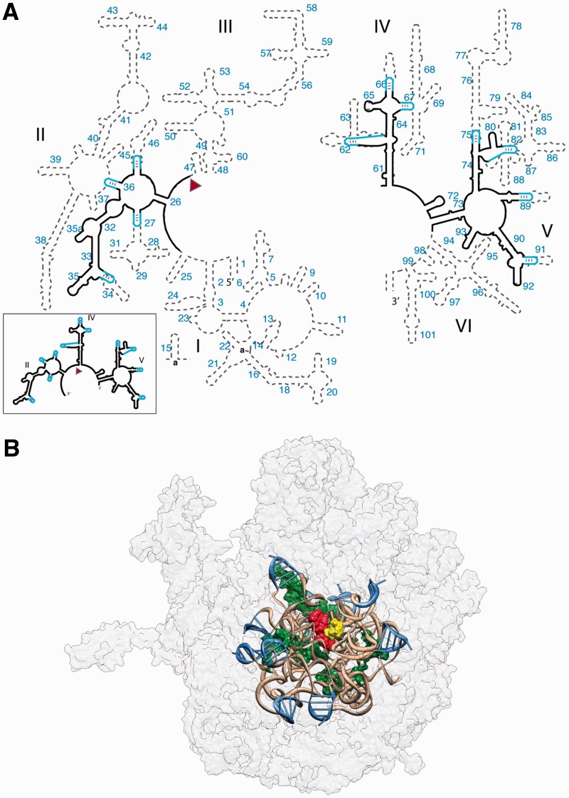
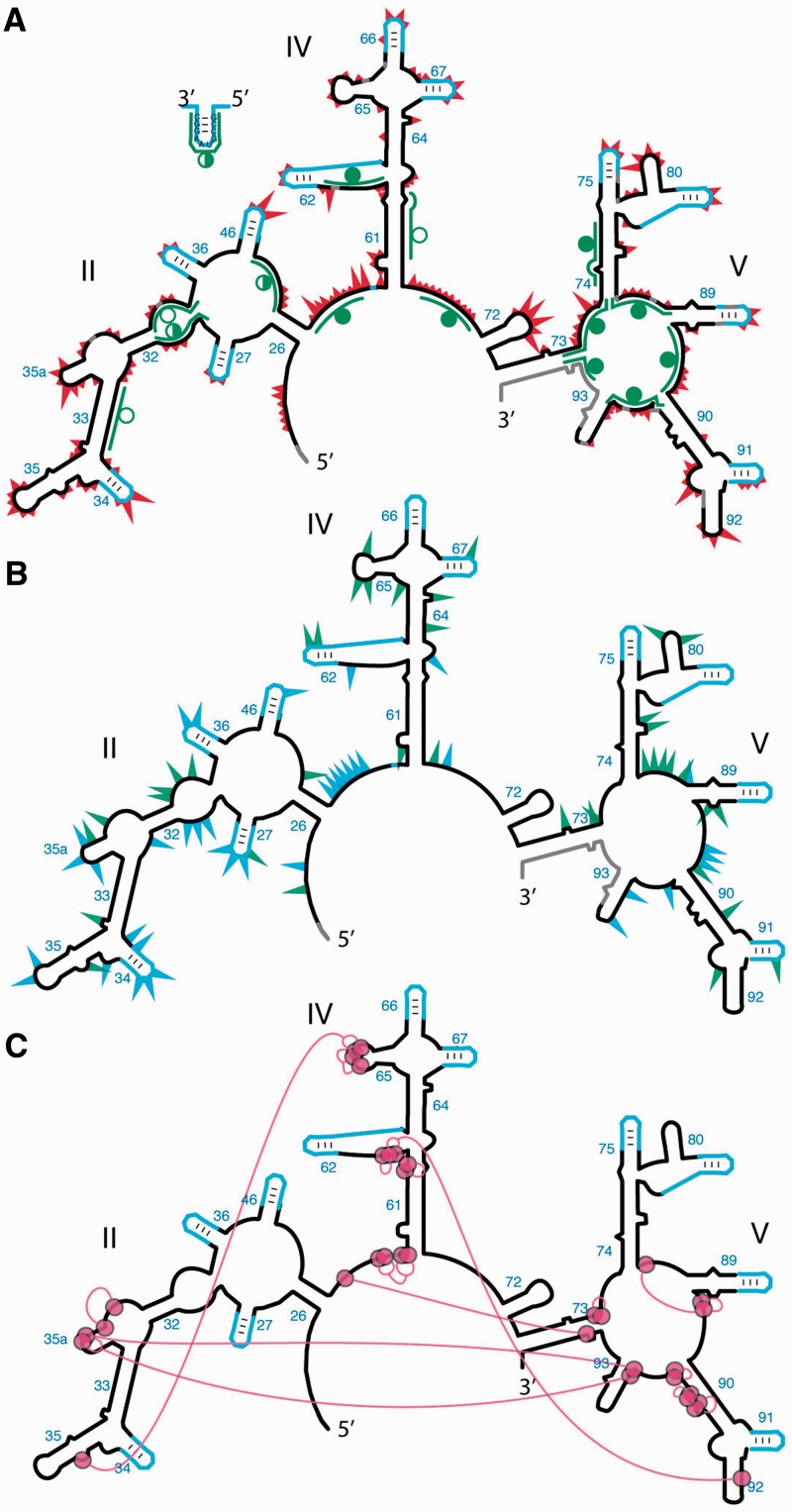
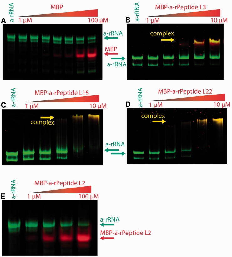
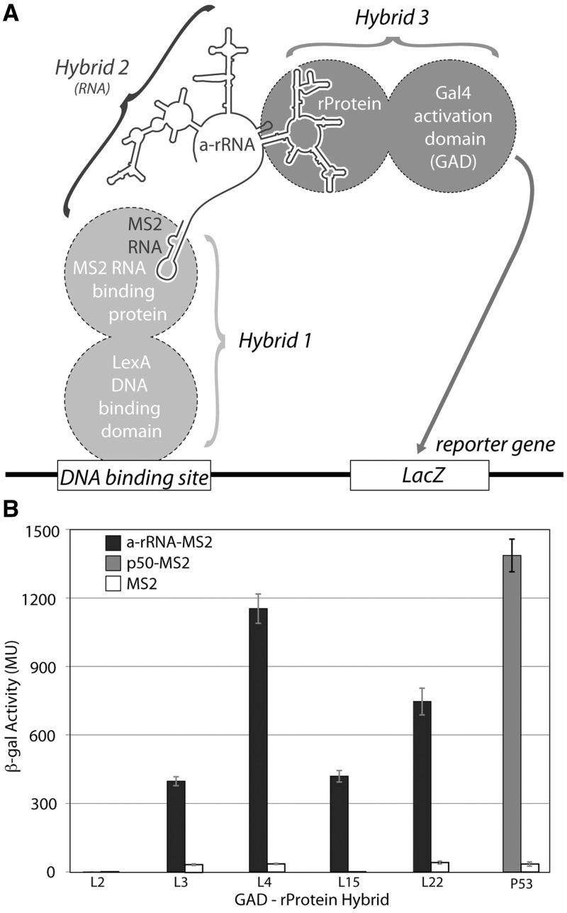
Ancient components of the ribosome, inferred from a consensus of previous work, were constructed in silico, in vitro and in vivo. The resulting model of the ancestral ribosome presented here incorporates ∼20% of the extant 23S rRNA and fragments of five ribosomal proteins. We test hypotheses that ancestral rRNA can: (i) assume canonical 23S rRNA-like secondary structure, (ii) assume canonical tertiary structure and (iii) form native complexes with ribosomal protein fragments. Footprinting experiments support formation of predicted secondary and tertiary structure. Gel shift, spectroscopic and yeast three-hybrid assays show specific interactions between ancestral rRNA and ribosomal protein fragments, independent of other, more recent, components of the ribosome. This robustness suggests that the catalytic core of the ribosome is an ancient construct that has survived billions of years of evolution without major changes in structure. Collectively, the data here support a model in which ancestors of the large and small subunits originated and evolved independently of each other, with autonomous functionalities.


Figures

Figure 1.
Figure 1.
Various models of 23S rRNA evolution. The dashed line illustrates the canonical secondary structure of the T. thermophilus 23S rRNA. Secondary structural domains are indicated by roman numerals. The red and green lines show the two inner shells of the ribosomal onion of Hsiao and Williams, marking the rRNA that is in closest proximity, in three dimensions, to the site of peptidyl transfer. The gray boxes are ancient according to the ‘A-minor’ method of Steinberg. The hashed boxes (with black horizontal lines) are ancient according to the networking analysis of Fox. Multidentate Mg2+-phosphate interactions, also proposed as an indicator of ancient rRNA, are indicated by magenta circles. The orange line shows the universally conserved portions of the 23S rRNA in bacteria, archaea, eukarya, and in mitochondria, as determined by Gutell and Harvey.
Figure 2.
Figure 2.
(A) Predicted secondary structure of the ancestral 23S rRNA. Ancestral fragments of rRNA, indicated by black lines in the secondary structure, are derived from a consensus of models of rRNA evolution. The ancestral rRNA elements are stitched together by stem loops (blue). The RNA sequences are from the T. thermophilus 23S rRNA. Helix numbers are indicated. The predicted secondary structure of the a-rRNA alone is highlighted in the outbox. (B) 3D model of the a-PTC. This 3D model contains the a-rRNA plus five a-rPeptides (ancestral fragments of ribosomal proteins L2, L3, L4, L15 and L22). a-rRNA is shown in ribbon (brown), the stem loops are blue and the peptides are in surface representation (green). For reference, A-site (yellow) and P-site (red) substrates are shown in the figure, but are not components of the a-PTC. The modern LSU surface is shown for comparison (light gray, transparent).
Figure 3.
Figure 3.
Probing the secondary and tertiary structure of a-rRNA. (A) SHAPE and RNase H mapping. Red triangles mark SHAPE reactivities in 250 mM Na+, mapped onto the predicted secondary structure of a-rRNA. Larger triangles indicate greater SHAPE reactivity. RNase H DNA probes are indicated by green lines. Circles indicate extent of RNA digestion by RNase H: filled circles (more than 75%), half-filled circles (between 25 and 75%) and empty circles (<25%). (B) Effects of 10 mM Mg2+ on SHAPE reactivity suggest formation of tertiary structure. Green triangles show the greatest increases in SHAPE reactivity upon addition of Mg2+. Blue triangles show the greatest decreases in reactivity. (C) Multidentate Mg2+-phosphate interactions observed in the T. thermophilus LSU (PDB 2J01) are mapped onto the predicted secondary structure of a-rRNA. Magenta circles indicate first-shell Mg2+-OP (magnesium-phosphate oxygen) interactions. Magenta lines indicate PO-Mg2+-OP linkages. Gray shading in panels A and B indicates rRNA where SHAPE data were not accessible. SHAPE reactions were performed in 50 mM NaHEPES, pH 8.0, 200 mM NaOAc, 0 or 10 mM MgCl2.
Figure 4.
Figure 4.
The effect of Mg2+ on gel mobility of the a-rRNA suggests Mg2+ induction of folding. Shown here is a-rRNA annealed in 10 mM Tris, pH 8.0, and varying [Mg2+], resolved on a 5% native acrylamide gel. Lane 1, [Mg2+] = 0 μM; Lane 2, 12.5; Lane 3, 25; Lane 4, 50; Lane 5, 100; Lane 6, 250; Lane 7, 500.
Figure 5.
Figure 5.
a-rRNA and a-rPeptide L4 are assembly competent and form a complex with 1:1 stoichiometry. Fluorescence signal at 334 nm is from the tryptophan residue of the a-rPeptide L4. In this plot of a continuous variation experiment of a-rPeptide L4 with a-rRNA, XL4 and Xa-rRNA values on horizontal axis denote mole fraction of the peptide and RNA in each sample, where the total concentration ([a-rRNA] + [a-rPeptide L4]) was held constant at 60 µM. The discontinuity at equivalent mole fractions of peptide and RNA indicates a complex with 1:1 stoichiometry. These binding assays were performed in 10 mM Tris, pH 8.0, at 25°C.
Figure 6.
Figure 6.
Gel shift analyses of interactions between a-rRNA and MBP-a-rPeptide fusions. RNA, protein and RNA–protein complexes were visualized on 5% native-PAGE gels by two-color EMSA. All binding reactions were performed with 1 µM a-rRNA, in 20 mM Tris buffer, pH 8.0. In each gel, the far left lanes contained a-rRNA only (no protein). (A) In a control assay, a-rRNA does not interact with MBP alone: a-rRNA incubated with MBP (left to right: 0, 1, 2, 4, 8, 10, 50 and 100 µM). (B) a-rRNA incubated with MBP-a-rPeptide L3 (left to right: 0, 1, 2, 4, 8 and 10 µM). (C) a-rRNA incubated with MBP-a-rPeptide L15 (left to right: 0, 1, 2, 4, 8 and 10 µM). (D) a-rRNA incubated with MBP-a-rPeptide L22 (left to right: 0, 1, 2, 4, 8 and 10 µM). (E) a-rRNA incubated with MBP-a-rPeptide L2 (left to right: 0, 1, 10, 50 and 100 µM).
Figure 7.
Figure 7.
(A) Schematic of the yeast three-hybrid assay used to characterize interactions between a-rRNA and ribosomal proteins. In yeast strain YBZ-1, the LacZ reporter gene is controlled by the LexA operator. Hybrid 1, a LexA/MS2 binding protein fusion, binds to the DNA binding site. The MS2 coat protein domain binds tightly to the MS2 RNA, which is fused to the RNA sequence of interest (e.g. a-rRNA). The rProtein of interest is fused to the yeast GAL4 transcriptional activation domain (GAD). In vivo RNA-protein binding results in expression of the LacZ reporter gene, which is quantified by β-gal activity. (B) In vivo interactions between a-rRNA and individual rProteins. Interaction is quantified by β-gal activity, reported in MU. Interactions were assayed between a-rRNA-MS2 and GAD-L2, GAD-L3, GAD-L4, GAD-L15 and GAD-L22 (black bars). Positive control was RNA aptamer p50-MS2 and GAD-p53 (gray bar). Negative controls consisted of MS2 RNA and the indicated protein hybrid (white bars).

Similar articles

Cited by

References

    1. Mears JA, Cannone JJ, Stagg SM, Gutell RR, Agrawal RK, Harvey SC. Modeling a minimal ribosome based on comparative sequence analysis. J. Mol. Biol. 2002;321:215–234. - PubMed
    1. Woese CR. Interpreting the universal phylogenetic tree. Proc. Natl. Acad. Sci. USA. 2000;97:8392–8396. - PMC - PubMed
    1. Woese CR. Translation: in retrospect and prospect. RNA. 2001;7:1055–1067. - PMC - PubMed
    1. Fournier GP, Neumann JE, Gogarten JP. Inferring the ancient history of the translation machinery and genetic code via recapitulation of ribosomal subunit assembly orders. PLoS One. 2010;5:e9437. - PMC - PubMed
    1. Williams D, Fournier GP, Lapierre P, Swithers KS, Green AG, Andam CP, Gogarten JP. A rooted net of life. Biol. Direct. 2011;6:45. - PMC - PubMed

Publication types

MeSH terms