. 2013 Mar 1;41(5):3314-26.
doi: 10.1093/nar/gkt019. Epub 2013 Jan 25.

MCPIP1 ribonuclease exhibits broad-spectrum antiviral effects through viral RNA binding and degradation

Affiliations

MCPIP1 ribonuclease exhibits broad-spectrum antiviral effects through viral RNA binding and degradation

Ren-Jye Lin et al. Nucleic Acids Res. .

Erratum in

Expression of concern in

Abstract

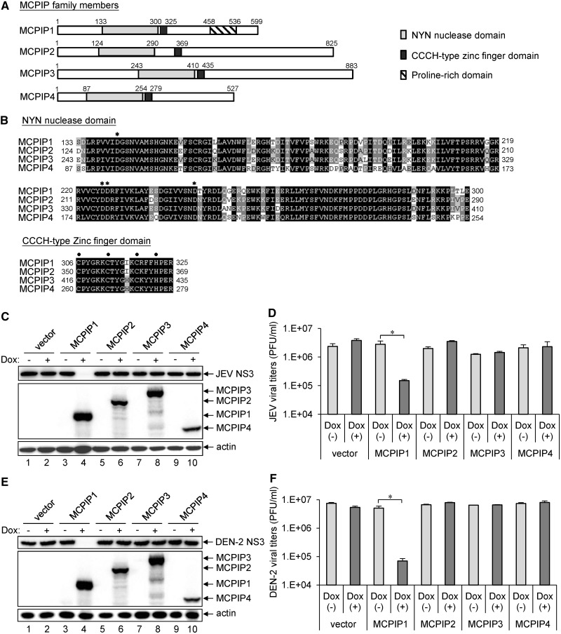
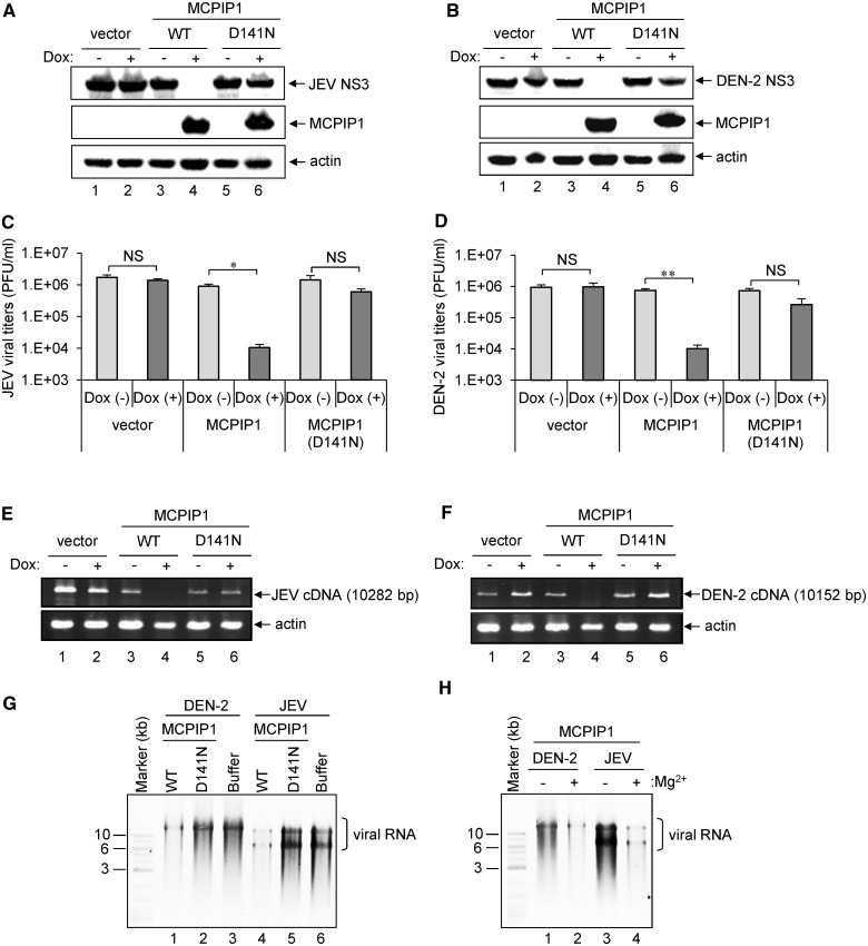
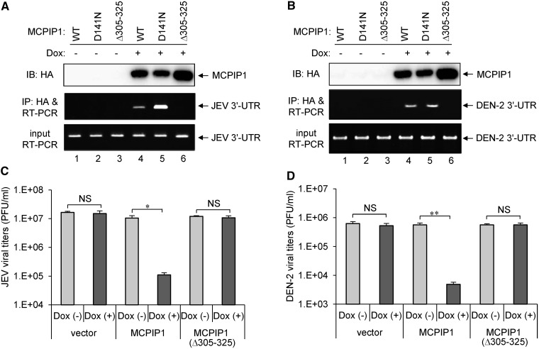
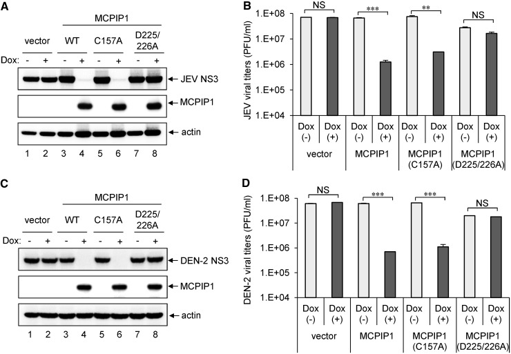
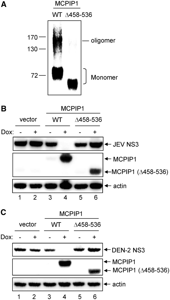
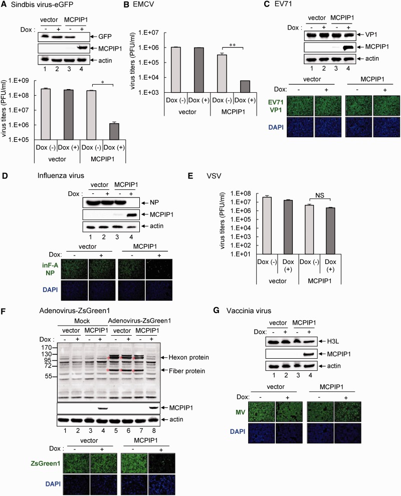
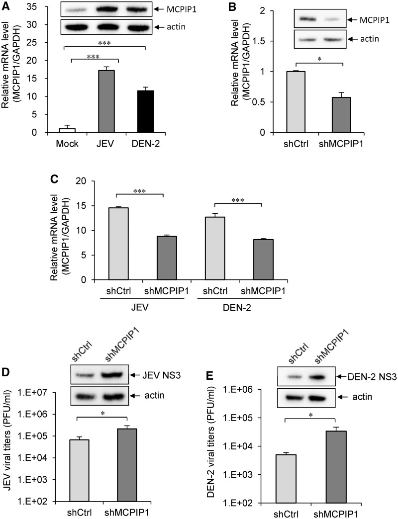
Monocyte chemoattractant protein 1-induced protein 1 (MCPIP1), belonging to the MCPIP family with highly conserved CCCH-type zinc finger and Nedd4-BP1, YacP Nuclease domains, has been implicated in negative regulation of the cellular inflammatory responses. In this report, we demonstrate for the first time that this RNA-binding nuclease also targets viral RNA and possesses potent antiviral activities. Overexpression of the human MCPIP1, but not MCPIP2, MCPIP3 or MCPIP4, inhibited Japanese encephalitis virus (JEV) and dengue virus (DEN) replication. The functional analysis of MCPIP1 revealed that the activities of RNase, RNA binding and oligomerization, but not deubiqutinase, are required for its antiviral potential. Furthermore, infection of other positive-sense RNA viruses, such as sindbis virus and encephalomyocarditis virus, and negative-sense RNA virus, such as influenza virus, as well as DNA virus, such as adenovirus, can also be blocked by MCPIP1. Moreover, the endogenous MCPIP1 gene expression was induced by JEV and DEN infection, and knockdown of MCPIP1 expression enhanced the replication of JEV and DEN in human cells. Thus, MCPIP1 can act as a host innate defense via RNase activity for targeting and degrading viral RNA.


Figures

Figure 1.
Figure 1.
The human MCPIP1 exhibits potent antiviral activity against JEV and DEN-2 infection. (A) Schematic representation of human MCPIP family proteins containing NYN and CCCH-type zinc finger domains. (B) Amino acid sequence alignment of NYN and CCCH-type zinc finger domains in human MCPIP family proteins. Identical amino acids are in bold and similar amino acids are in grey. Four conserved negative-charged Asp (D) residues in the NYN domain are indicated by asterisks. The three Cys and one His residues in the CCCH-type zinc finger domain are indicated by dots. The human T-REx-293 cells stably transfected with pcDNA5/TO vector or pcDNA5/TO with MCPIP1/2/3/4 were cultured in medium without (−) or with (+) Dox (1 µg/ml) for 12 h, and then cells were infected with JEV or DEN-2 (MOI 5) for 24 h. (C, E) Western blot analysis of protein expression of HA for MCPIP, JEV NS3, DEN-2 NS3 and actin. (D, F) Viral titration was done by plaque-forming assay on BHK-21 cells. Virus titers are shown as means and standard deviations of two independent experiments. The data of the indicated groups were compared by two-tailed Student’s t-tests. *P ≤ 0.05.
Figure 2.
Figure 2.
RNase activity of human MCPIP1 is essential for its antiviral effect against JEV and DEN-2 infection. (A, B) Human T-REx-293 cells with inducible expression of vector, MCPIP1 or MCPIP1-D141N mutant were cultured in medium without (−) or with (+) Dox (1 µg/ml) for 12 h, then infected with JEV or DEN-2 (MOI 5). At 24 h post-infection, cell lysates were harvested for western blot analysis of viral protein NS3 expression for JEV (A) and DEN-2 (B). Virus titers of JEV (C) and DEN-2 (D) in the culture supernatants were determined by plaque-forming assays on BHK-21 cells. Results are means and standard deviations of two independent experiments. Data were compared by two-tailed Student’s t-tests. *P ≤ 0.05; **P ≤ 0.01; NS: not significant. The viral RNA levels of JEV (E) and DEN-2 (F) were analysed by RT-PCR as described in ‘Materials and Methods’ section. The expected sizes of JEV and DEN-2 PCR products are shown on the right. (G) HA-tagged wild-type (WT) or MCPIP1-D141N mutant proteins were incubated with in vitro transcribed full-length DEN-2 or JEV RNA in reaction buffer with 5 mM Mg2+ at 30°C for 1 h. RNA was separated by 0.8% agarose gel electrophoresis and stained with ethidium bromide. The photos are shown as inverse images. (H) In vitro cleavage of human MCPIP1 degrading viral RNA was analysed without (−) or with (+) Mg2+.
Figure 3.
Figure 3.
The CCCH-type zinc finger domain of human MCPIP1 is required for viral RNA binding and antiviral activity against JEV and DEN-2 infection. (A, B) Human T-REx-293 cells with WT or MCPIP1-D141N or −Δ305 to 325 mutant were infected with JEV or DEN-2 (MOI 5) for 18 h, then cultured in medium without (−) or with (+) Dox (1 µg/ml) for 18 h. The viral RNA bound with MCPIP1 protein was pulled down with HA-beads and amplified by RT-PCR with JEV or DEN-2 3′-UTR specific primers (middle panels). RT-PCR of mRNA levels of JEV or DEN-2 3′-UTR in virus-infected cells (bottom panels). Western blot analysis of HA-tagged MCPIP1 protein expression in cell lysates (top panels). (C, D) Human T-REx-293 cells with vector, MCPIP1 or MCPIP1-Δ305–325 mutant were cultured without (−) or with (+) Dox (1 µg/ml) for 12 h and then infected with JEV or DEN-2 (MOI 5) for 24 h. Virus titers of JEV (C) and DEN-2 (D) were determined by plaque-forming assays on BHK-21 cells. Results are means and standard deviations of two independent experiments. The titers of the indicated groups were compared by two-tailed Student’s t-tests. *P ≤ 0.05; **P ≤ 0.01; NS: not significant.
Figure 4.
Figure 4.
Deubiqutinase activity of human MCPIP1 is not involved in its antiviral potential against JEV and DEN-2 infection. Human T-REx-293 cells with vector, WT or MCPIP1-C157A or -D225/226A were cultured without (−) or with (+) Dox (1 µg/ml) for 12 h, then infected with JEV or DEN-2 (MOI 5) for 24 h. Western blot analysis of levels of indicated proteins (A, C). Virus titers of JEV (B) and DEN-2 (D) are means and standard deviations of two independent experiments. The titers of the indicated groups were compared by two-tailed Student’s t-tests. **P ≤ 0.01; ***P ≤ 0.001; NS: not significant.
Figure 5.
Figure 5.
The proline-rich domain of human MCPIP1 is essential for its oligomerization and antiviral activity. (A) Human T-REx-293 cells with WT or MCPIP1-Δ458–536 mutant were cultured with Dox (1 µg/ml) for 24 h. The cell proteins were cross-linked with disuccinimidyl suberate, and HA-tagged MCPIP1 proteins were pulled down with HA-beads. The oligomerization of MCPIP1 was analysed by SDS-PAGE and immunoblotted with anti-HA antibody. (B, C) Human T-REx-293 cells with vector, WT or MCPIP1-Δ458–536 mutant were cultured without (−) or with (+) Dox (1 µg/ml) for 12 h and then infected with JEV or DEN-2 (MOI 5) for 24 h. The cell lysates were harvested for western blotting with the indicated antibodies.
Figure 6.
Figure 6.
The antiviral potential of human MCPIP1 against several viruses. Human T-REx-293 cells with MCPIP1 or vector control were cultured without (−) or with (+) Dox (1 µg/ml) for 12 h and then infected with the indicated viruses for 24 h. (A) Western blot analysis of the indicated proteins in cells with sindbis-eGFP infection (MOI 20) (upper panel). Virus titers of sindbis-eGFP were determined by plaque-forming assays on BHK-21 cells (lower panel). (B) For EMCV infection (MOI 1), virus titers were determined by plaque-forming assays on Vero cells. (C) Western blot analysis of levels of indicated proteins with EV71 infection (MOI 5) (upper panels). Immunofluorescence assay of EV71 viral protein VP1 (green) and DAPI (blue) was photographed by fluorescent microscopy (lower panels). (D) Western blot analysis of levels of indicated proteins to detect influenza virus infection (MOI 1) (upper panels). Immunofluorescence assay of influenza viral protein NP (green) and DAPI (blue) (lower panels). (E) For VSV infection (MOI 0.01), virus titers were determined by plaque-forming assays on Vero cells. (F) Western blot analysis of the protein expression of adenovirus hexon and fiber proteins to detect adenovirus-ZsGreen1 infection (MOI 20) (upper panels). Immunofluorescence assay of adenovirus expression of ZsGreen1 (green) and DAPI (blue) (lower panels). (G) Western blot analysis of level of vaccinia viral protein H3L to detect VV infection (MOI 5) (upper panels). Immunofluorescence assay of mature vaccinia virus (MV) (green) and DAPI (blue) (lower panels). The results are means and standard deviations of two independent experiments. The viral titers were compared by two-tailed Student’s t-tests. *P ≤ 0.05; **P ≤ 0.01. NS: not significant.
Figure 7.
Figure 7.
The antiviral potential of endogenous MCPIP1. (A) The human A549 cells were mock infected or infected with JEV (MOI 5 for 24 h) or DEN-2 (MOI 5 for 48 h). The relative MCPIP1 mRNA levels were analysed by quantitative real-time RT-PCR and normalized to that of GAPDH mRNA. The MCPIP1 protein levels were assessed by western blotting with MCPIP1-specific antibody. (B) A549 cells were transduced with MCPIP shRNA lentiviral particles (shMCPIP1) or control shRNA lentiviral particles (shCtrl). The knockdown effect was evaluated by real-time RT-PCR and western blotting as described in panel A. (C) The MCPIP1 expression levels in A549-shCtrl and A549-shMCPIP1 cells infected with JEV (MOI 5 for 24 h) or DEN-2 (MOI 5 for 48 h) were analysed by real-time RT-PCR. A549-shCtrl and A549-shMCPIP1 cells infected with (D) JEV (MOI 5 for 24 h) or (E) DEN-2 (MOI 5 for 48 h) were analysed for viral production by plaque-forming assay and viral protein expression by western blotting. Data are shown as means and standard deviations of three independent experiments. The data of the indicated groups were compared by two-tailed Student’s t-tests. *P ≤ 0.05; ***P ≤ 0.001.

References

    1. Bick MJ, Carroll JW, Gao G, Goff SP, Rice CM, MacDonald MR. Expression of the zinc-finger antiviral protein inhibits alphavirus replication. J. Virol. 2003;77:11555–11562. - PMC - PubMed
    1. Gao G, Guo X, Goff SP. Inhibition of retroviral RNA production by ZAP, a CCCH-type zinc finger protein. Science. 2002;297:1703–1706. - PubMed
    1. Muller S, Moller P, Bick MJ, Wurr S, Becker S, Gunther S, Kummerer BM. Inhibition of filovirus replication by the zinc finger antiviral protein. J. Virol. 2007;81:2391–2400. - PMC - PubMed
    1. Brown RS. Zinc finger proteins: getting a grip on RNA. Curr. Opin. Struct. Biol. 2005;15:94–98. - PubMed
    1. Hall TM. Multiple modes of RNA recognition by zinc finger proteins. Curr. Opin. Struct. Biol. 2005;15:367–373. - PubMed

Publication types

MeSH terms