

Topical niacinamide 4% and desonide 0.05% for treatment of axillary hyperpigmentation: a randomized, double-blind, placebo-controlled study
- PMID: 23355788
- PMCID: PMC3552481
- DOI: 10.2147/CCID.S39246
Topical niacinamide 4% and desonide 0.05% for treatment of axillary hyperpigmentation: a randomized, double-blind, placebo-controlled study
Abstract
Background: Axillary hyperpigmentation is a frequent cause of cosmetic consultations in dark-skinned women from tropical areas, including Latin America. Currently, there is no widely accepted treatment for the disorder, but it is usually treated with bleaching agents because it is considered a variant of inflammatory hyperpigmentation. The purpose of this study was to assess the efficacy of niacinamide 4% and desonide 0.05% emulsions compared with placebo in the treatment of axillary hyperpigmentation.
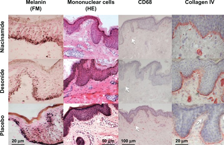
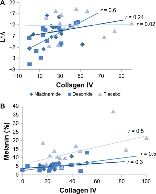
Methods: Twenty-four women aged 19-27 years with hyperpigmented axillae (phototype III-V) were randomly assigned to receive the study treatments in the axillary region. Improvement was assessed at baseline, then clinically and by colorimetry 9 weeks later. Quantitative evaluation including melanin, inflammatory infiltrates, NKI/Beteb, CD1a, CD68, and collagen type IV content was performed by histochemistry and immunohistochemistry, assisted by computerized morphometric analysis.
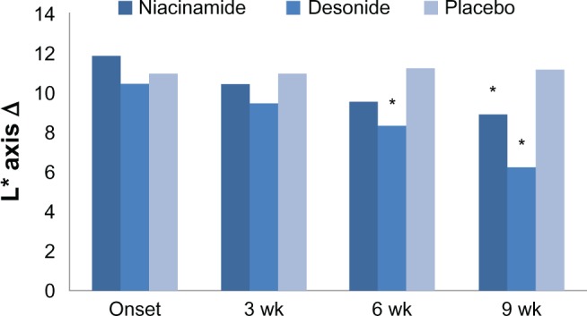
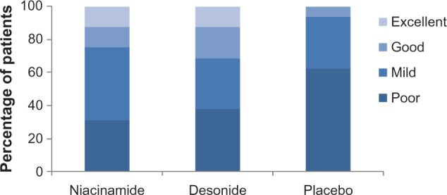
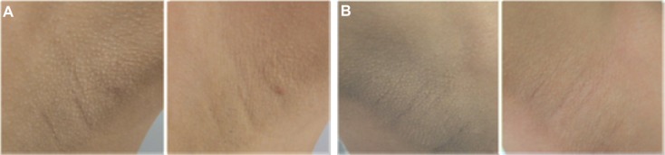
Results: Both niacinamide and desonide induced significant colorimetric improvement compared with placebo; however, desonide showed a better depigmenting effect than niacinamide. A good to excellent response was achieved in 24% of cases for niacinamide, 30% for desonide, and 6% for placebo. We observed a marked disruption of the basal membrane in axillary hyperpigmentation and an inflammatory infiltrate that improved after treatment. Decreased pigmentation in the desonide-treated axillae was associated with recovery of disruption at the basal membrane.
Conclusion: Niacinamide and desonide showed depigmenting properties in women with axillary hyperpigmentation. These findings may be explained by their antimelanogenic and anti-inflammatory properties, respectively.
Keywords: desonide; niacinamide; post-inflammatory hyperpigmentation.
Figures
References
-
- Alexis AF, Sergay AB, Taylor SC. Common dermatologic disorders in skin of color: a comparative practice survey. Cutis. 2007;80:387–394. - PubMed
-
- Papa CM, Kligman AM. The behavior of melanocytes in inflammation. J Invest Dermatol. 1965;45:465–473. - PubMed
-
- Mottaz JH, Thorne EG, Zelickson AS. Response of the epidermal melanocyte to minor trauma. Arch Dermatol. 1971;104:611–618. - PubMed
-
- Tomita Y, Maeda K, Tagami H. Melanocyte-stimulating properties of arachidonic acid metabolites: possible role in postinflammatory pigmentation. Pigment Cell Res. 1992;5:357–361. - PubMed
-
- Lamel SA, Rahvar M, Maibach HI. Postinflammatory hyperpigmentation secondary to external insult: an overview of the quantitative analysis of pigmentation. Cutan Ocul Toxicol. 2012;32:67–71. - PubMed
LinkOut - more resources
Full Text Sources
Other Literature Sources
