

DNA damage and transcriptional changes in the gills of mytilus galloprovincialis exposed to nanomolar doses of combined metal salts (Cd, Cu, Hg)
- PMID: 23355883
- PMCID: PMC3552849
- DOI: 10.1371/journal.pone.0054602
DNA damage and transcriptional changes in the gills of mytilus galloprovincialis exposed to nanomolar doses of combined metal salts (Cd, Cu, Hg)
Abstract
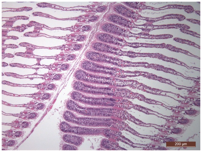
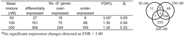
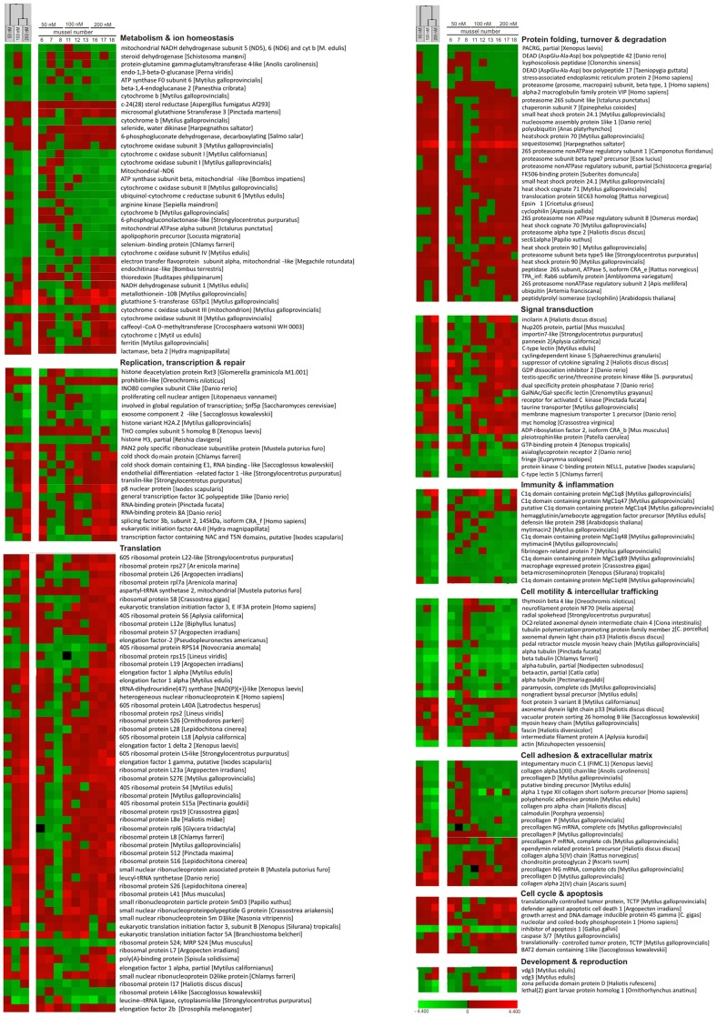
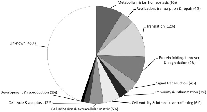
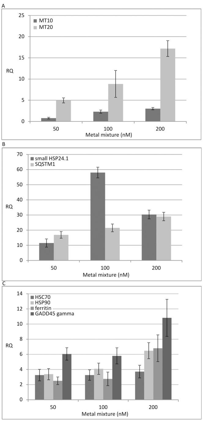
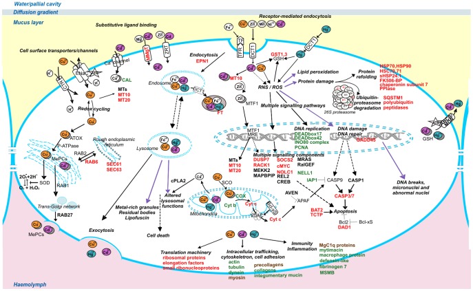
Aiming at an integrated and mechanistic view of the early biological effects of selected metals in the marine sentinel organism Mytilus galloprovincialis, we exposed mussels for 48 hours to 50, 100 and 200 nM solutions of equimolar Cd, Cu and Hg salts and measured cytological and molecular biomarkers in parallel. Focusing on the mussel gills, first target of toxic water contaminants and actively proliferating tissue, we detected significant dose-related increases of cells with micronuclei and other nuclear abnormalities in the treated mussels, with differences in the bioconcentration of the three metals determined in the mussel flesh by atomic absorption spectrometry. Gene expression profiles, determined in the same individual gills in parallel, revealed some transcriptional changes at the 50 nM dose, and substantial increases of differentially expressed genes at the 100 and 200 nM doses, with roughly similar amounts of up- and down-regulated genes. The functional annotation of gill transcripts with consistent expression trends and significantly altered at least in one dose point disclosed the complexity of the induced cell response. The most evident transcriptional changes concerned protein synthesis and turnover, ion homeostasis, cell cycle regulation and apoptosis, and intracellular trafficking (transcript sequences denoting heat shock proteins, metal binding thioneins, sequestosome 1 and proteasome subunits, and GADD45 exemplify up-regulated genes while transcript sequences denoting actin, tubulins and the apoptosis inhibitor 1 exemplify down-regulated genes). Overall, nanomolar doses of co-occurring free metal ions have induced significant structural and functional changes in the mussel gills: the intensity of response to the stimulus measured in laboratory supports the additional validation of molecular markers of metal exposure to be used in Mussel Watch programs.
Conflict of interest statement
Figures

Similar articles
-
Assessment of low-level metal contamination using the Mediterranean mussel gills as the indicator tissue.Environ Sci Pollut Res Int. 2010 May;17(4):977-86. doi: 10.1007/s11356-009-0270-x. Epub 2009 Dec 9. Environ Sci Pollut Res Int. 2010. PMID: 20012221
-
Transcriptional response of the mussel Mytilus galloprovincialis (Lam.) following exposure to heat stress and copper.PLoS One. 2013 Jun 25;8(6):e66802. doi: 10.1371/journal.pone.0066802. Print 2013. PLoS One. 2013. PMID: 23825565 Free PMC article.
-
Proteomic response of mussels Mytilus galloprovincialis exposed to CuO NPs and Cu²⁺: an exploratory biomarker discovery.Aquat Toxicol. 2014 Oct;155:327-36. doi: 10.1016/j.aquatox.2014.07.015. Epub 2014 Jul 21. Aquat Toxicol. 2014. PMID: 25089921
-
Measurement of p-nitrophenyl acetate esterase activity (EA), total antioxidant capacity (TAC), total oxidant status (TOS) and acetylcholinesterase (AChE) in gills and digestive gland of Mytilus galloprovincialis exposed to binary mixtures of Pb, Cd and Cu.Environ Sci Pollut Res Int. 2016 Dec;23(24):25385-25392. doi: 10.1007/s11356-016-7677-y. Epub 2016 Oct 1. Environ Sci Pollut Res Int. 2016. PMID: 27696195
-
Comparative study of the accumulation and detoxification of Cu (essential metal) and Hg (nonessential metal) in the digestive gland and gills of mussels Mytilus galloprovincialis, using analytical and histochemical techniques.Chemosphere. 2011 May;83(8):1155-65. doi: 10.1016/j.chemosphere.2011.01.003. Epub 2011 Feb 1. Chemosphere. 2011. PMID: 21288554
Cited by
-
Spermatozoa Transcriptional Response and Alterations in PL Proteins Properties after Exposure of Mytilus galloprovincialis to Mercury.Int J Mol Sci. 2021 Feb 5;22(4):1618. doi: 10.3390/ijms22041618. Int J Mol Sci. 2021. PMID: 33562685 Free PMC article.
-
Altered Gene Expression in the Schistosome-Transmitting Snail Biomphalaria glabrata following Exposure to Niclosamide, the Active Ingredient in the Widely Used Molluscicide Bayluscide.PLoS Negl Trop Dis. 2015 Oct 9;9(10):e0004131. doi: 10.1371/journal.pntd.0004131. eCollection 2015. PLoS Negl Trop Dis. 2015. PMID: 26452273 Free PMC article.
-
The contents of heavy metals (cd, cr, as, pb, ni, and sn) in the selected commercial yam powder products in South Korea.Prev Nutr Food Sci. 2013 Dec;18(4):249-55. doi: 10.3746/pnf.2013.18.4.249. Prev Nutr Food Sci. 2013. PMID: 24551826 Free PMC article.
-
Integration of small RNAs, degradome and transcriptome sequencing in hyperaccumulator Sedum alfredii uncovers a complex regulatory network and provides insights into cadmium phytoremediation.Plant Biotechnol J. 2016 Jun;14(6):1470-83. doi: 10.1111/pbi.12512. Epub 2016 Jan 23. Plant Biotechnol J. 2016. PMID: 26801211 Free PMC article.
-
Transcriptional responses of Biomphalaria pfeifferi and Schistosoma mansoni following exposure to niclosamide, with evidence for a synergistic effect on snails following exposure to both stressors.PLoS Negl Trop Dis. 2019 Dec 16;13(12):e0006927. doi: 10.1371/journal.pntd.0006927. eCollection 2019 Dec. PLoS Negl Trop Dis. 2019. PMID: 31841501 Free PMC article.
References
-
- Goldberg ED (1975) The Mussel Watch - a first step in global marine monitoring. Mar Pollut Bull 6: 111.
-
- Nguyen TT, Hayes BJ, Guthridge K, Ab Rahim ES, Ingram BA (2011) Use of a microsatellite-based pedigree in estimation of heritabilities for economic traits in Australian blue mussel, Mytilus galloprovincialis . J Anim Breed Genet 128(6): 482–90. - PubMed
-
- Lu Q, Hwang DS, Liu Y, Zeng H (2012) Molecular interactions of mussel protective coating protein, mcfp-1, from Mytilus californianus . Biomaterials 33(6): 1903–11. - PubMed
-
- Fields PA, Zuzow MJ, Tomanek L (2012) Proteomic responses of blue mussel (Mytilus) congeners to temperature acclimation. J Exp Biol 215(Pt 7): 1106–16. - PubMed
Publication types
MeSH terms
Substances
LinkOut - more resources
Full Text Sources
Other Literature Sources
Medical
Molecular Biology Databases
