. 2013;8(1):e54754.
doi: 10.1371/journal.pone.0054754. Epub 2013 Jan 23.

In vivo cell reprogramming towards pluripotency by virus-free overexpression of defined factors

Affiliations

In vivo cell reprogramming towards pluripotency by virus-free overexpression of defined factors

Açelya Yilmazer et al. PLoS One. 2013.

Abstract

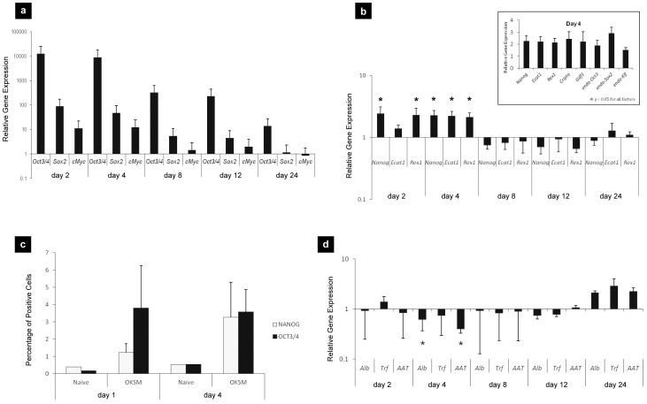
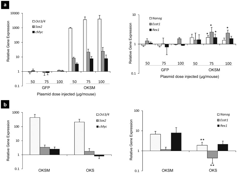
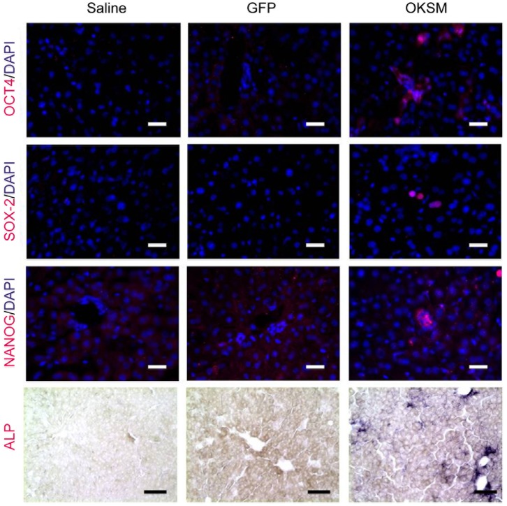
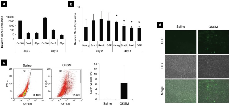
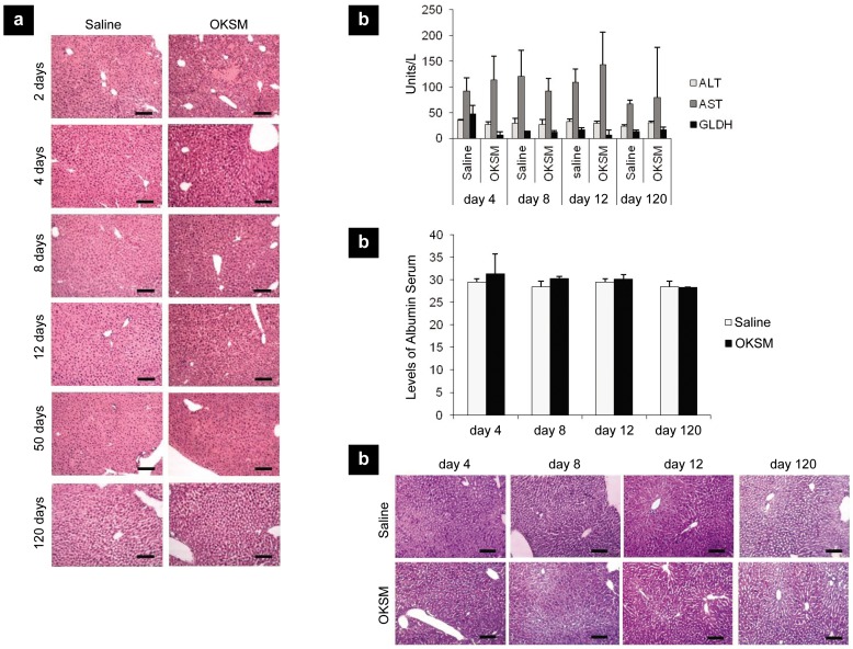
The ability to induce the reprogramming of somatic mammalian cells to a pluripotent state by the forced expression of specific transcription factors has helped redefine the rules of cell fate and plasticity, as well as open possibilities for disease modeling, drug screening and regenerative medicine. Here, we hypothesized that the non-viral forced expression of the four originally discovered defined factors (OKSM) in adult mice could result in in vivo reprogramming of cells in the transfected tissue in situ. We show that a single hydrodynamic tail-vein (HTV) injection of two plasmids encoding for Oct3/4, Sox2, Klf4 and c-Myc respectively, are highly expressed in the liver tissue of Balb/C adult mice. Hallmark pluripotency markers were upregulated within 24-48 h after injection, followed by down-regulation of all major hepatocellular markers. Generation of transcriptionally reprogrammed cells in vivo was further confirmed by positive staining of liver tissue sections for all major pluripotency markers in Balb/C mice and the Nanog-GFP reporter transgenic strain (TNG-A) with concomitant upregulation of GFP expression in situ. No signs of physiological or anatomical abnormalities or teratoma formation were observed in the liver examined up to 120 days. These findings indicate that virus-free expression of OKSM factors in vivo can transcriptionally reprogram cells in situ rapidly, efficiently and transiently, absent of host tissue damage or teratoma formation.


Conflict of interest statement

Competing Interests: The authors have declared that no competing interests exist.

Figures

Figure 1
Figure 1. In vivo overexpression of OKSM transcription factors in adult mouse liver.
Balb/C mice were HTV injected with 0.9% saline alone, 75 µg of pCX-OKS-2A and 75 µg pCX-cMyc in 0.9% saline and at days 2, 4, 8, 12, 24, RT-qPCR analysis of hepatocytes was performed to determine the relative gene expression of: (a) transfected transcription factors (OKSM) and (b) endogenous pluripotency markers. All gene expression levels were normalized to HTV-injected saline group (*p<0.05 indicates statistically significant differences between the expression levels of pluripotency markers in the OKSM and saline HTV-injected groups, obtained by the analysis of variance and Tukey's pairwise comparison); (c) flow cytometry analysis of OCT3/4 positive and NANOG positive cells in liver extracts; (d) relative gene expression of hepatocyte markers as determined by RT-qPCR. All gene expression levels were normalized to saline HTV-injected group (* p<0.05 indicates statistically significant differences between the expression levels for hepatocyte markers in the OKSM and saline HTV-injected groups, obtained by the analysis of variance and Tukey's pairwise comparison).
Figure 2
Figure 2. OKSM and OKS factor overexpression with dose-response in adult mouse liver.
Balb/C mice HTV injected with 0.9% saline alone, pCX-OKS-2A with (OSKM) and without (OKS) pCX-cMyc in 0.9% saline, or pCAG-GFP in 0.9% saline, at the indicated doses. On day 4, RT-qPCR analysis of hepatocyte extracts was performed. (a) Expression levels of the injected reprogramming transcription factors and endogenous pluripotency genes were determined for plasmid dose-escalation (total plasmid dose 50, 75 and 100 µg/animal). All gene expression levels were normalized to the HTV-injected saline group (*p<0.05 indicates statistically significant differences between the expression levels of pluripotency markers in the OKSM and saline HTV-injected groups, obtained by the analysis of variance and Tukey's pairwise comparison); (b) Expression levels of the injected reprogramming transcription factors and endogenous pluripotency genes with and without inclusion of cMyc. All gene expression levels were normalized to HTV-injected saline group (**p<0.01 indicates statistically significant differences between the expression levels of pluripotency markers in the OKSM and OKS injected groups, obtained by the analysis of variance and Tukey's pairwise comparison).
Figure 3
Figure 3. In vivo cell reprogramming on adult mouse liver tissue by immunohistochemistry.
Balb/C mice HTV injected with 0.9% saline alone, 75 µg of pCX-OKS-2A and 75 µg pCX-cMyc in 0.9% saline, or 150 µg of pCAG-GFP in 0.9% saline and at day 4, livers were collected and frozen tissue sections were stained with anti-OCT4, anti-SOX2 or anti-NANOG antibodies to assess immunoreactivity, or BCIP/NBT to determine ALP activity in the tissue (40x). Scale bars represent 100 µm.
Figure 4
Figure 4. In vivo cell reprogramming in TNG-A mice.
TNG-A mice were HTV injected with 0.9% saline alone, 75 µg of pCX-OKS-2A and 75 µg pCX-cMyc in 0.9% saline and at days 2, 4, RT-qPCR analysis of hepatocytes was performed to determine the relative gene expression of: (a) transfected transcription factors (OKSM) and (b) endogenous pluripotency markers. All gene expression levels were normalized to HTV-injected saline group (*p<0.05 indicates statistically significant differences between the expression levels of pluripotency markers in the OKSM and saline HTV-injected groups, obtained by the analysis of variance and Tukey's pairwise comparison); (c) flow cytometry analysis of GFP positive cells in liver extracts; (d) liver tissue frozen and sectioned to image GFP-positive cells with fluorescence microscopy at day 4 (10x).
Figure 5
Figure 5. The effect of in vivo cell reprogramming on hepatotoxicity and liver damage.
Balb/C mice HTV injected with either 75 µg of pCX-OKS-2A and 75 µg pCX-cMyc in 0.9% saline or 0.9% saline only. On days 2, 4, 8, 12, 50 and 120 livers and sera were isolated. (a) H&E staining of liver sections; (b) levels of liver enzymes and (c) albumin were analyzed; (d) liver sections were PAS stained to determine glycogen storage levels. Representative images were captured with light microscopy (10x). Scale bars represent 100 µm.

References

    1. Takahashi K, Tanabe K, Ohnuki M, Narita M, Ichisaka T, et al. (2007) Induction of Pluripotent Stem Cells from Adult Human Fibroblasts by Defined Factors. Cell 131: 861–872. - PubMed
    1. Yu J, Vodyanik MA, Smuga-Otto K, Antosiewicz-Bourget J, Frane JL, et al. (2007) Induced Pluripotent Stem Cell Lines Derived from Human Somatic Cells. Science 318: 1917–1920. - PubMed
    1. Stadtfeld M, Nagaya M, Utikal J, Weir G, Hochedlinger K (2008) Induced Pluripotent Stem Cells Generated Without Viral Integration. Science 322: 945–949. - PMC - PubMed
    1. Okita K, Nakagawa M, Hyenjong H, Ichisaka T, Yamanaka S (2008) Generation of Mouse Induced Pluripotent Stem Cells Without Viral Vectors. Science 322: 949–953. - PubMed
    1. Gonzalez F, Barragan Monasterio M, Tiscornia G, Montserrat Pulido N, Vassena R, et al. (2009) Generation of mouse-induced pluripotent stem cells by transient expression of a single nonviral polycistronic vector. Proc Natl Acad Sci USA 106: 8918–8922. - PMC - PubMed

Publication types