. 2013;8(1):e54926.
doi: 10.1371/journal.pone.0054926. Epub 2013 Jan 23.

Oxygen tension modulates differentiation and primary macrophage functions in the human monocytic THP-1 cell line

Affiliations

Oxygen tension modulates differentiation and primary macrophage functions in the human monocytic THP-1 cell line

Ana Cristina G Grodzki et al. PLoS One. 2013.

Abstract

The human THP-1 cell line is widely used as an in vitro model system for studying macrophage differentiation and function. Conventional culture conditions for these cells consist of ambient oxygen pressure (∼20% v/v) and medium supplemented with the thiol 2-mercaptoethanol (2-ME) and serum. In consideration of the redox activities of O₂ and 2-ME, and the extensive experimental evidence supporting a role for reactive oxygen species (ROS) in the differentiation and function of macrophages, we addressed the question of whether culturing THP-1 cells under a more physiologically relevant oxygen tension (5% O₂) in the absence of 2-ME and serum would alter THP-1 cell physiology. Comparisons of cultures maintained in 18% O₂versus 5% O₂ indicated that reducing oxygen tension had no effect on the proliferation of undifferentiated THP-1 cells. However, decreasing the oxygen tension to 5% O₂ significantly increased the rate of phorbol ester-induced differentiation of THP-1 cells into macrophage-like cells as well as the metabolic activity of both undifferentiated and PMA-differentiated THP-1 cells. Removal of both 2-ME and serum from the medium decreased the proliferation of undifferentiated THP-1 cells but increased metabolic activity and the rate of differentiation under either oxygen tension. In differentiated THP-1 cells, lowering the oxygen tension to 5% O₂ decreased phagocytic activity, the constitutive release of β-hexosaminidase and LPS-induced NF-κB activation but enhanced LPS-stimulated release of cytokines. Collectively, these data demonstrate that oxygen tension influences THP-1 cell differentiation and primary macrophage functions, and suggest that culturing these cells under tightly regulated oxygen tension in the absence of exogenous reducing agent and serum is likely to provide a physiologically relevant baseline from which to study the role of the local redox environment in regulating THP-1 cell physiology.


Conflict of interest statement

Competing Interests: The authors have declared that no competing interests exist.

Figures

Figure 1
Figure 1. Influence of O2 tension, 2-ME and serum on proliferation of undifferentiated THP-1 cells.
Monocytic THP-1 cells were synchronized by serum deprivation for 48 h and then cultured in hyperoxic (18% O2) or normoxic (5% O2) with or without 2-ME and/or FBS. Cell density was determined using a hemocytometer at 24 h (A) and 48 h (B) after synchronization. Data are presented as the mean ± SEM (n = 5 independent experiments). *Significantly different from cultures with 2-ME and FBS under the same oxygen tension at p<0.05 (one-way ANOVA with post hoc Tukey’s test).
Figure 2
Figure 2. Influence of O2 tension, 2-ME and serum on the metabolic activity of THP-1 cells.
THP-1 cells were synchronized by serum deprivation for 48 h. (A) Undifferentiated THP-1 cells were plated in 96-well plates precoated with poly-D-lysine. (B) THP-1 cells were triggered to undergo macrophage differentiation by incubating with PMA at 20 ng/ml for 24 h. MTT was added to both undifferentiated and differentiated THP-1 cells for 3 h under varying O2, 2-ME and serum conditions. MTT reduction, measured as the absorbance at 562 nm, was normalized to protein concentration. Data are presented as the mean ± SEM (n = 4 independent experiments. *Significantly different from +2-ME+FBS (standard culture conditions) under the same oxygen tension (one-way ANOVA and post hoc Tukey’s test); Significantly different from –2-ME+FBS under the same oxygen tension (one-way ANOVA and post hoc Tukey’s test); #Significantly different from the same culture condition in the 18% O2 group (e.g., 18% O2 versus 5% O2) by Student’s t-test. *, #, p<0.05; **,##, ▴▴ p<0.01; ***, ###, ▴▴▴ p<0.001.
Figure 3
Figure 3. Influence of O2 tension, 2-ME and serum on the rate of THP-1 differentiation.
Differentiation of THP-1 cells from monocytic to macrophage cells is associated with transition from a non-adherent to an adherent cell type. To determine whether culture conditions affect PMA-stimulated differentiation of THP-1 cells to macrophages, cell adhesion was assessed at 3 and 24 h after addition of PMA (20 ng/ml) to the culture medium. Data are presented as the mean ± SEM of the protein concentration of adherent cells at 3 h as a percentage of the protein concentration of adherent cells at 24 h (n = 4 independent experiments). *Significantly different from +2-ME+FBS (standard culture conditions) under the same oxygen tension (one-way ANOVA and post hoc Tukey’s test); Significantly different from –2-ME+FBS under the same oxygen tension (one-way ANOVA and post hoc Tukey’s test); #Significantly different from the same culture condition in the 18% O2 group (e.g., 18% O2 versus 5% O2) by Student’s t-test. **, ##, ▴▴ p<0.01; ***, ###, ▴▴▴ p<0.001.
Figure 4
Figure 4. Influence of O2 tension, 2-ME and serum on release of β-hexosaminidase.
Differentiated THP-1 cells constitutively release β-hexosaminidase that is measurable in the conditioned medium (supernatant) after 24 h (A) or 48 h (B) of culture. β-Hexosaminidase is also detected in cell lysates (C). β-Hexosaminidase activity per well was normalized to the concentration of protein in the same well as determined using the BCA protein assay. Data are presented as mean ± SEM (n = 3 independent experiments). *Significantly different from +2-ME+FBS (standard culture conditions) under the same oxygen tension (one-way ANOVA and post hoc Tukey’s test); Significantly different from –2-ME+FBS under the same oxygen tension (one-way ANOVA and post hoc Tukey’s test); #Significantly different from the same culture condition in the 18% O2 group (e.g., 18% O2 versus 5% O2) by Student’s t-test. *, #, p<0.05; **, ##, ▴▴ p<0.01; ***, ###, ▴▴▴ p<0.001.
Figure 5
Figure 5. Oxygen tension significantly influences phagocytosis in PMA-differentiated THP-1 cells.
Undifferentiated THP-1 cells were synchronized by serum deprivation for 48 h, plated at a density of 105cells/well in a 96-well plate and differentiated with PMA (20 ng/ml) for 48 h in the absence of 2-ME and FBS. Differentiated THP-1 cells were washed and then incubated for 3 h with E.coli BioParticles®, which emit fluorescence upon acidification in lysosomes following phagocytosis. Phagocytosis, which was quantified by determining the fluorescence intensity at 600 nm, was blocked by pretreating cultures with cytochalasin D (2 µM) for 1 h prior to addition of E. coli BioParticles®. The mean fluorescence intensity was normalized to protein concentration as determined using the BCA protein assay. Data are presented as the mean ± SEM (n = 3 independent experiments). *Significantly different from control (– cytochalasin) treatment under the same oxygen tension; #significantly different from the same culture condition in the 18% O2 treatment group (e.g., 18% O2 versus 5% O2) by Student’s t-test. ***, ### p<0.001.
Figure 6
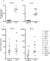
Figure 6. Oxygen tension influences LPS-induced NF-κB activation and release of cytokines in PMA-differentiated THP-1 cells.
Undifferentiated THP-1 XBlue cells, which express an NF-κB reporter gene linked to secreted embryonic alkaline phosphatase (SEAP) were synchronized by serum deprivation for 48 h, and then differentiated with PMA (20 ng/ml) for 48 h in the absence of 2-ME and FBS. Differentiated THP-1 XBlue cells were then cultured in the absence (baseline) or presence of LPS (1 µg/ml) for an additional 24 h in either 18% (A, C) or 5% (B, D) O2. SEAP activity was quantified by QuantiBlue at 630 nm (A,B). Conditioned media from these cultures were analyzed using a human Milliplex Kit® to simultaneously quantify multiple cytokines and chemokines released from differentiated THP-1 cell during the 24 h incubation. Each symbol in panels C and D represents the mean of duplicates from one of five wells run in a representative experiment. Data are presented as the mean ± SEM (n = 2 independent experiments). ***Significantly different from baseline under the same oxygen tension at p<0.001, ###significantly different from 18% O2 at p<0.001 (Student’s t-test).
Figure 7
Figure 7. Oxygen tension influences redox in LPS-induced NF-κB activation in PMA-differentiated THP-1 cells.
Undifferentiated THP-1 XBlue cells, which express an NF-κB reporter gene linked to secreted embryonic alkaline phosphatase (SEAP) were synchronized by serum deprivation for 48 h, and then differentiated with PMA (20 ng/ml) for 48 h. Differentiated THP-1 XBlue cells were then cultured in varying concentrations of DPI (A) or NGA (B) for 4 h followed by LPS (1 µg/ml) stimulation for an additional 24 h in either 18% or 5% O2. SEAP activity was quantified by QuantiBlue at 630 nm. Data are presented as the mean ± SEM (n = 2 independent experiments). *Significantly different from baseline (SEAP activity in the absence of inhibitor) at the same oxygen tension; **p<0.01; ***p<0.001 (one-way ANOVA with post hoc Tukey’s test). #Significantly different from 5% O2 at same antioxidant concentration; # p<0.05; ## p<0.01 (Student’s t-test).

Similar articles

Cited by

  • The Role of Tissue Oxygen Tension in Dengue Virus Replication.
    Frakolaki E, Kaimou P, Moraiti M, Kalliampakou KI, Karampetsou K, Dotsika E, Liakos P, Vassilacopoulou D, Mavromara P, Bartenschlager R, Vassilaki N. Frakolaki E, et al. Cells. 2018 Dec 1;7(12):241. doi: 10.3390/cells7120241. Cells. 2018. PMID: 30513781 Free PMC article.
  • Guidelines for the use and interpretation of assays for monitoring autophagy (4th edition)1.
    Klionsky DJ, Abdel-Aziz AK, Abdelfatah S, Abdellatif M, Abdoli A, Abel S, Abeliovich H, Abildgaard MH, Abudu YP, Acevedo-Arozena A, Adamopoulos IE, Adeli K, Adolph TE, Adornetto A, Aflaki E, Agam G, Agarwal A, Aggarwal BB, Agnello M, Agostinis P, Agrewala JN, Agrotis A, Aguilar PV, Ahmad ST, Ahmed ZM, Ahumada-Castro U, Aits S, Aizawa S, Akkoc Y, Akoumianaki T, Akpinar HA, Al-Abd AM, Al-Akra L, Al-Gharaibeh A, Alaoui-Jamali MA, Alberti S, Alcocer-Gómez E, Alessandri C, Ali M, Alim Al-Bari MA, Aliwaini S, Alizadeh J, Almacellas E, Almasan A, Alonso A, Alonso GD, Altan-Bonnet N, Altieri DC, Álvarez ÉMC, Alves S, Alves da Costa C, Alzaharna MM, Amadio M, Amantini C, Amaral C, Ambrosio S, Amer AO, Ammanathan V, An Z, Andersen SU, Andrabi SA, Andrade-Silva M, Andres AM, Angelini S, Ann D, Anozie UC, Ansari MY, Antas P, Antebi A, Antón Z, Anwar T, Apetoh L, Apostolova N, Araki T, Araki Y, Arasaki K, Araújo WL, Araya J, Arden C, Arévalo MA, Arguelles S, Arias E, Arikkath J, Arimoto H, Ariosa AR, Armstrong-James D, Arnauné-Pelloquin L, Aroca A, Arroyo DS, Arsov I, Artero R, Asaro DML, Aschner M, Ashrafizadeh M, Ashur-Fabian O, Atanasov AG, Au AK, Auberger P, Auner HW, Aurelian L, Autelli R… See abstract for full author list ➔ Klionsky DJ, et al. Autophagy. 2021 Jan;17(1):1-382. doi: 10.1080/15548627.2020.1797280. Epub 2021 Feb 8. Autophagy. 2021. PMID: 33634751 Free PMC article.
  • Immunostimulant Bathing Influences the Expression of Immune- and Metabolic-Related Genes in Atlantic Salmon Alevins.
    Figueiredo F, Kristoffersen H, Bhat S, Zhang Z, Godfroid J, Peruzzi S, Præbel K, Dalmo RA, Xu X. Figueiredo F, et al. Biology (Basel). 2021 Sep 29;10(10):980. doi: 10.3390/biology10100980. Biology (Basel). 2021. PMID: 34681079 Free PMC article.
  • An improved organotypic cell culture system to study tissue-resident macrophages ex vivo.
    Aktories P, Petry P, Glatz P, Andrieux G, Oschwald A, Botterer H, Gorka O, Erny D, Boerries M, Henneke P, Groß O, Prinz M, Kierdorf K. Aktories P, et al. Cell Rep Methods. 2022 Jul 26;2(8):100260. doi: 10.1016/j.crmeth.2022.100260. eCollection 2022 Aug 22. Cell Rep Methods. 2022. PMID: 36046625 Free PMC article.
  • Hydrogen peroxide-induced oxidative stress and its impact on innate immune responses in lung carcinoma A549 cells.
    Upadhyay S, Vaish S, Dhiman M. Upadhyay S, et al. Mol Cell Biochem. 2019 Jan;450(1-2):135-147. doi: 10.1007/s11010-018-3380-2. Epub 2018 Jun 25. Mol Cell Biochem. 2019. PMID: 29938378

References

    1. Tsuchiya S, Yamabe M, Yamaguchi Y, Kobayashi Y, Konno T, et al. (1980) Establishment and characterization of a human acute monocytic leukemia cell line (THP-1). Int J Cancer 26: 171–176. - PubMed
    1. Csete M (2005) Oxygen in the cultivation of stem cells. Ann N Y Acad Sci 1049: 1–8. - PubMed
    1. Yan HM, Ramachandran A, Bajt ML, Lemasters JJ, Jaeschke H (2010) The oxygen tension modulates acetaminophen-induced mitochondrial oxidant stress and cell injury in cultured hepatocytes. Toxicol Sci 117: 515–523. - PMC - PubMed
    1. Lu SC (2009) Regulation of glutathione synthesis. Mol Aspects Med 30: 42–59. - PMC - PubMed
    1. Meister A, Anderson ME (1983) Glutathione. Annu Rev Biochem 52: 711–760. - PubMed

Publication types

MeSH terms