. 2011;1(2):142-163.
doi: 10.2174/2210296511101020142.

Patents on Technologies of Human Tissue and Organ Regeneration from Pluripotent Human Embryonic Stem Cells

Patents on Technologies of Human Tissue and Organ Regeneration from Pluripotent Human Embryonic Stem Cells

Xuejun H Parsons et al. Recent Pat Regen Med. 2011.

Abstract

Human embryonic stem cells (hESCs) are genetically stable with unlimited expansion ability and unrestricted plasticity, proffering a pluripotent reservoir for in vitro derivation of a large supply of disease-targeted human somatic cells that are restricted to the lineage in need of repair. There is a large healthcare need to develop hESC-based therapeutic solutions to provide optimal regeneration and reconstruction treatment options for the damaged or lost tissue or organ that have been lacking. In spite of controversy surrounding the ownership of hESCs, the number of patent applications related to hESCs is growing rapidly. This review gives an overview of different patent applications on technologies of derivation, maintenance, differentiation, and manipulation of hESCs for therapies. Many of the published patent applications have been based on previously established methods in the animal systems and multi-lineage inclination of pluripotent cells through spontaneous germ-layer differentiation. Innovative human stem cell technologies that are safe and effective for human tissue and organ regeneration in the clinical setting remain to be developed. Our overall view on the current patent situation of hESC technologies suggests a trend towards hESC patent filings on novel therapeutic strategies of direct control and modulation of hESC pluripotent fate, particularly in a 3-dimensional context, when deriving clinically-relevant lineages for regenerative therapies.


Figures
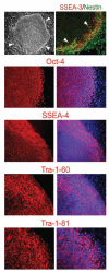
Fig. (1)
Fig. (1)
bFGF is a critical component that sustains undifferentiated growth of hESCs on human feeders. Phase contrast image shows the highly compact undifferentiated morphology of a hESC colony on human feeder cells. White arrows delineate the edge of a hESC colony. Immunofluorescence images show that hESCs inside the colonies express the undifferentiated hESC markers Oct-4 (red), SSEA-4 (red), Tra-1-60 (red), and Tra-1-81 (red). Cells at the edge of the colonies exhibit the classic flattened epithelial morphology indicative of the onset of differentiation, and express SSEA-3 (red) and Nestin (green). Cells that have migrated outside the colonies continued to differentiate into large elliptoid-appearing cells that persist in expressing Nestin, but cease expressing SSEA-3, Oct-4, SSEA-4, Tra-1-60, and Tra-1-81. All cells are revealed by DAPI staining of their nuclei (blue) in the merged images.
Fig. (2)
Fig. (2)
bFGF is a critical component that sustains undifferentiated growth of hESCs on laminin/collagen (Matrigel). Phase contrast images show the highly compact undifferentiated morphology of a hESC colony on laminin/collagen. White arrows delineate the edge of a hESC colony. Immunofluorescence images show that hESCs inside the colonies express the undifferentiated hESC markers Oct-4 (red), SSEA-4 (red), Tra-1-60 (red), and Tra-1-81 (red). Cells at the edge of the colonies exhibit the classic flattened epithelial morphology indicative of the onset of differentiation, and express SSEA-3 (red) and Nestin (green). Cells that have migrated outside the colonies continued to differentiate into large elliptoid-appearing cells that persist in expressing Nestin and Vimentin (red), but cease expressing SSEA-3, Oct-4, SSEA-4, Tra-1-60, and Tra-1-81. All cells are revealed by DAPI staining of their nuclei (blue) in the merged images. bFGF short-term proliferation assay shows that, without bFGF or with a low concentration of bFGF (4 ng/ml), hESCs displayed significantly slow growth rates. With bFGF at a concentration ranging from 10 to 50ng/ml, hESCs displayed a comparable growth rate as those maintained in MEF-conditioned media (CM). bFGF dose-response assay shows that hESCs maintained in media containing 20 ng/ml bFGF exhibited the highest undifferentiated percentage.
Fig. (3)
Fig. (3)
bFGF, insulin, ascorbic acid, and laminin are minimal essential requirements for the maintenance of undifferentiated hESCs. Quantitative analysis of defined components with hESCs seeded on purified human laminin and cultivated in a base medium indicates that bFGF, insulin, and ascorbic acid are essential components for maintaining substantial numbers of hESCs in undifferentiated state, indicated by Oct-4 positive. With all the components or in the absence of transferrin, a majority of hESC colonies displayed a highly compact undifferentiated morphology and expressed Oct-4 (red). In the absence of albumin, hESC colonies were more flat and spread out (white square delineates the same area shown in the inset), but a large proportion continued to express Oct-4 and exhibited a highly compact morphology. However, if ascorbic acid was omitted from the media (NO Ascorbic Acid), the colonies often became very dense in the center and cyst-like (red arrows). Undifferentiated hESCs maintained in media containing both bFGF and insulin do not express SSEA-1 (red). Absence of either bFGF or insulin induces complete differentiation, as indicated by SSEA-1 expression. Large round cells were usually present in media that contained only insulin, and elliptically-shaped cells were present in media that contained only bFGF. Absence of ascorbic acid (NO Ascorbic Acid) resulted in slower cell growth in media containing only insulin, but accelerated the differentiated growth in the dense centers of colonies in media containing only bFGF (red arrows). White arrows delineate the edge of hESC colonies. All cells are indicated by DAPI staining of their nuclei (blue).
Fig. (4)
Fig. (4)
(A) Other growth factors can not replace bFGF for the maintenance of undifferentiated hESCs. hESC colonies maintained in aFGF, EGF, IGF-I, IGF-II, PDGF, VEGF, activin-A, and BMP-2 generally display a more differentiated morphology that consists of dense centers containing cyst-like structures and cells heaping upon each other (red arrows). Note that, although most cells are differentiated, a minority of the small colonies (<30%) retain a compact morphology (blue arrows) and continue to express Oct-4 (red). White square indicates the approximate area that is visualized at higher magnification in the right. DAPI staining is blue. (B) Determining the minimal essential matrix. hESCs maintained on laminin have a classic undifferentiated morphology and express Oct-4 (red). DAPI staining of their nuclei is blue. White arrows delineate the edge of a hESC colony. In contrast, hESC colonies maintained on fibronectin, collagen IV, or gelatin displayed a more differentiated morphology within their first passage.
Fig. (5)
Fig. (5)
Retinoic acid (RA) signals neural induction direct of pluripotence under defined conditions. (A) Upon exposure of hESCs to RA under the defined culture system, large differentiated Oct-4 (red) negative cells within the colony began to emerge. RA-induced differentiated Oct-4-negative cells began to express SSEA-1 (red), HNK1 (red), and AP2 (red), consistent with early neuroectodermal differentiation. These cells continued to mature ultimately expressing the neuronal marker Map-2 (green), usually in areas where cells began to pile up. All cells are indicated by DAPI staining of their nuclei (blue). White arrows delineate the edge of a hESC colony. (B) The induced hESCs formed cardioblasts (Nkx2.5+, with nicotinamide [NAM], see Fig. 6) or neuroblasts (β-III-tubulin+, with RA) in suspension, as compared to germlayer-induced embryoid bodies (EBs) derived from hESCs without treatment (Control). (C) RA treatment induces differentiation towards a neuronal lineage with a drastic increase in efficiency, as assessed by the percentages of cells that expressed β-III-tubulin (red) and coexpressed Map-2 (green) (arrow, pigmented cells). (D) Nurr1 translocates to the nucleus upon exposure of hESCs to RA. A large subpopulation of these hESC-derived neuronal cells progressed to express tyrosine hydroxylase (TH, red) in the presence sonic hedgehog (+Shh) or absence Shh (–Shh). A subpopulation of these hESC-derived Map-2-positive (green) cells began to express Hb9 (red) and Lim3 (red) (shown in a 3D matrix). All cells are indicated by DAPI nuclear staining (blue).
Fig. (6)
Fig. (6)
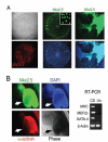
Nicotinamide (NAM) signals cardiac induction direct of pluripotence under defined conditions. (A) Upon exposure of hESCs to NAM under the defined culture system, large differentiated Oct-4 (red) negative cells within the colony began to emerge. NAM-induced Oct- 4-negative cells began to express SSEA-1 (red), Nkx2.5 (green), consistent with early cardiac differentiation. Progressively increased intensity of Nkx2.5 was usually observed in areas of the colony where cells began to pile up. (B) NAM-induced hESCs yielded beating cardiomyocytes with a drastic increase in efficiency, as assessed by the percentages of cellular clusters that displayed rhythmic contractions (arrows), and immunopositive for Nkx2.5 (green) and α-actinin (red), and expressed cardiomyocyte markers cardiac MHC, MEF2c, and GATA-4 (CD), as compared to undifferentiated hESCs as the control (Un). All cells are indicated by DAPI nuclear staining (blue).

Similar articles

Cited by

References

    1. Thomson JA, Itskovitz-Eldor J, Shapiro SS, Waknitz MA, Swiergiel JJ, Marshall VS, et al. Embryonic stem cell lines derived from human blastocysts. Science. 1998;282:1145–7. - PubMed
    1. Parsons XH, Teng YD, Snyder EY. Important precautions when deriving patient-specific neural elements from pluripotent cells. Cytotherapy. 2009;11:815–24. - PMC - PubMed
    1. Kirby T. Robert Edwards: Nobel Prize for father of in-vitro fertilization. Lancet. 2010;376:1293–0. - PubMed
    1. Martino G, Pluchino S. The therapeutic potential of neural stem cells. Nat Rev. 2006;7:395–406. - PubMed
    1. Redmond DE, Jr, Bjugstad KB, Teng YD, Ourednik V, Ourednik J, Wakeman DR, et al. Behavioral improvement in a primate Parkinson's model is associated with multiple homeostatic effects of human neural stem cells. Proc Natl Acad Sci USA. 2007;104:12175–80. - PMC - PubMed

LinkOut - more resources