Changes in red blood cell membrane structure in type 2 diabetes: a scanning electron and atomic force microscopy study
- PMID: 23356738
- PMCID: PMC3599682
- DOI: 10.1186/1475-2840-12-25
Changes in red blood cell membrane structure in type 2 diabetes: a scanning electron and atomic force microscopy study
Abstract
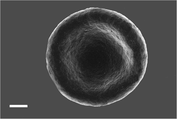
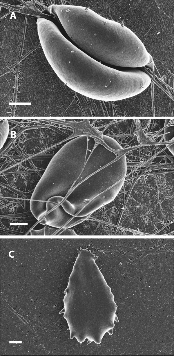
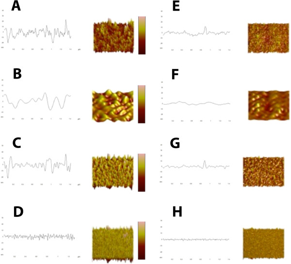
Red blood cells (RBCs) are highly deformable and possess a robust membrane that can withstand shear force. Previous research showed that in diabetic patients, there is a changed RBC ultrastructure, where these cells are elongated and twist around spontaneously formed fibrin fibers. These changes may impact erythrocyte function. Ultrastructural analysis of RBCs in inflammatory and degenerative diseases can no longer be ignored and should form a fundamental research tool in clinical studies. Consequently, we investigated the membrane roughness and ultrastructural changes in type 2 diabetes. Atomic force microscopy (AFM) was used to study membrane roughness and we correlate this with scanning electron microscopy (SEM) to compare results of both the techniques with the RBCs of healthy individuals. We show that the combined AFM and SEM analyses of RBCs give valuable information about the disease status of patients with diabetes. Effectiveness of treatment regimes on the integrity, cell shape and roughness of RBCs may be tracked, as this cell's health status is crucial to the overall wellness of the diabetic patient.
Figures


Similar articles
-
Topological features of erythrocytes in thalassemic patients: quantitative characterization by scanning electron and atomic force microscopy.Anal Quant Cytopathol Histpathol. 2014 Apr;36(2):91-9. Anal Quant Cytopathol Histpathol. 2014. PMID: 24902361
-
Changes in red blood cell membrane structure in G6PD deficiency: an atomic force microscopy study.Clin Chim Acta. 2015 Apr 15;444:264-70. doi: 10.1016/j.cca.2015.02.042. Epub 2015 Mar 4. Clin Chim Acta. 2015. PMID: 25748035
-
Characterizing pathology in erythrocytes using morphological and biophysical membrane properties: Relation to impaired hemorheology and cardiovascular function in rheumatoid arthritis.Biochim Biophys Acta Biomembr. 2017 Dec;1859(12):2381-2391. doi: 10.1016/j.bbamem.2017.09.014. Epub 2017 Sep 15. Biochim Biophys Acta Biomembr. 2017. PMID: 28919343
-
Structure and function in native and pathological erythrocytes: a quantitative view from the nanoscale.Micron. 2012 Dec;43(12):1273-86. doi: 10.1016/j.micron.2012.03.019. Epub 2012 Apr 3. Micron. 2012. PMID: 22537716 Review.
-
Nanoscale Surface Characterization of Human Erythrocytes by Atomic Force Microscopy: A Critical Review.IEEE Trans Nanobioscience. 2015 Sep;14(6):625-33. doi: 10.1109/TNB.2015.2424674. Epub 2015 Apr 28. IEEE Trans Nanobioscience. 2015. PMID: 25935044 Review.
Cited by
-
Poorly controlled type 2 diabetes is accompanied by significant morphological and ultrastructural changes in both erythrocytes and in thrombin-generated fibrin: implications for diagnostics.Cardiovasc Diabetol. 2015 Mar 8;14:30. doi: 10.1186/s12933-015-0192-5. Cardiovasc Diabetol. 2015. PMID: 25848817 Free PMC article.
-
Beyond the myocardium? SGLT2 inhibitors target peripheral components of reduced oxygen flux in the diabetic patient with heart failure with preserved ejection fraction.Heart Fail Rev. 2022 Jan;27(1):219-234. doi: 10.1007/s10741-020-09996-y. Heart Fail Rev. 2022. PMID: 32583230 Review.
-
Membrane Lesions and Reduced Life Span of Red Blood Cells in Preeclampsia as Evidenced by Atomic Force Microscopy.Int J Mol Sci. 2023 Apr 12;24(8):7100. doi: 10.3390/ijms24087100. Int J Mol Sci. 2023. PMID: 37108270 Free PMC article.
-
Morphology, membrane nanostructure and stiffness for quality assessment of packed red blood cells.Sci Rep. 2017 Aug 10;7(1):7846. doi: 10.1038/s41598-017-08255-9. Sci Rep. 2017. PMID: 28798476 Free PMC article.
-
Searching for the Mechanical Fingerprint of Pre-diabetes in T1DM: A Case Report Study.Front Bioeng Biotechnol. 2020 Sep 29;8:569978. doi: 10.3389/fbioe.2020.569978. eCollection 2020. Front Bioeng Biotechnol. 2020. PMID: 33117782 Free PMC article.
References
-
- Kozlova EK, Chernysh AM, Moroz VV, Kuzovlev AN. Analysis of nanostructure of red blood cells membranes by space Fourier transform of AFM images. Micron. 2012;44:218–227. - PubMed
-
- Girasole M, Pompeo G, Cricenti A, Congiu-Castellano A, Andreola F, Serafino A, Frazer BH, Boumis G, Amiconi G. Roughness of the plasma membrane as an independent morphological parameter to study RBCs: a quantitative atomic force microscopy investigation. Biochim Biophys Acta. 2007;1768(5):1268–1276. doi: 10.1016/j.bbamem.2007.01.014. - DOI - PubMed
MeSH terms
LinkOut - more resources
Full Text Sources
Other Literature Sources
Medical
Miscellaneous