Review
. 2013 May 13;425(9):1461-75.
doi: 10.1016/j.jmb.2013.01.027. Epub 2013 Jan 25.

The energy and work of a ligand-gated ion channel

Affiliations
Review

The energy and work of a ligand-gated ion channel

Anthony Auerbach. J Mol Biol. .

Abstract

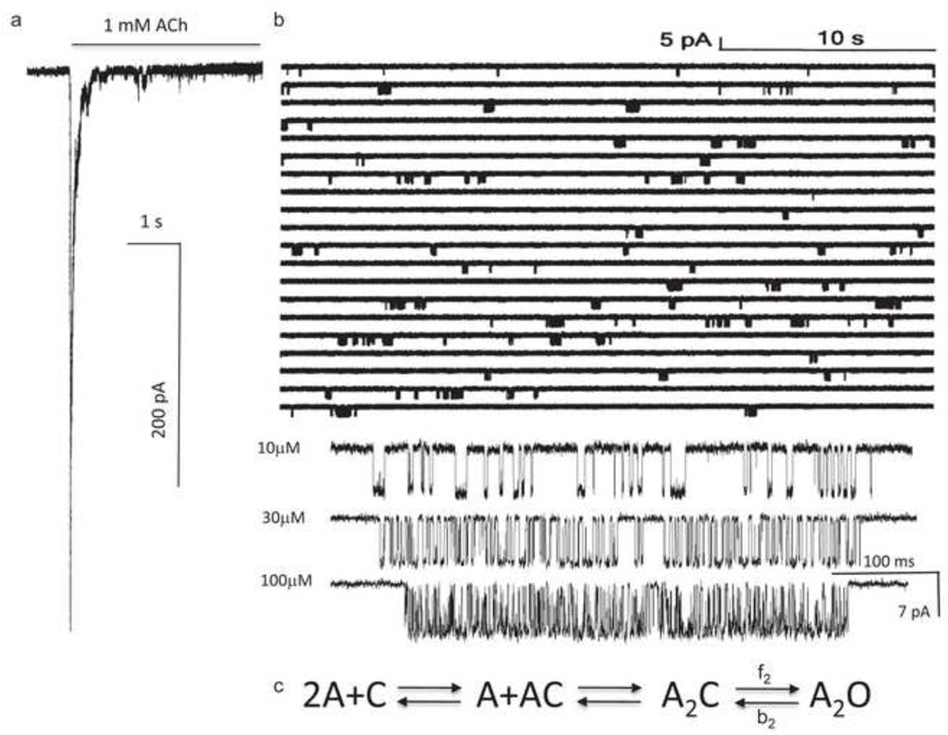
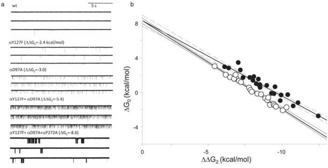
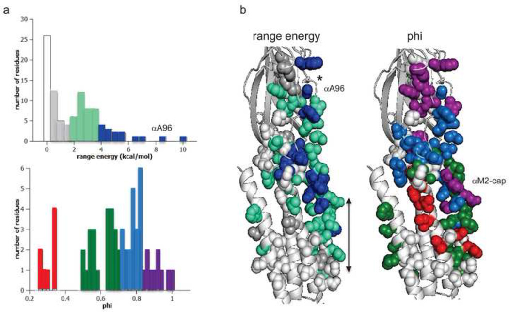
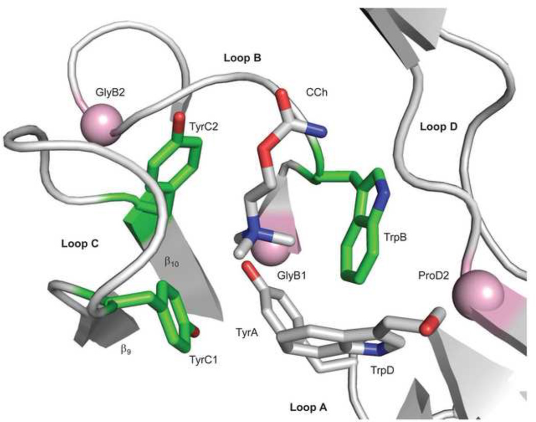
Ligand-gated ion channels are allosteric membrane proteins that isomerize between C(losed) and O(pen) conformations. A difference in affinity for ligands in the two states influences the C↔O "gating" equilibrium constant. The energies associated with adult-type mouse neuromuscular nicotinic acetylcholine receptor (AChR) channel gating have been measured by using single-channel electrophysiology. Without ligands, the free energy, enthalpy and entropy of gating are ΔG0=+8.4, ΔH0=+10.9 and TΔS0=+2.5kcal/mol (-100mV, 23°C). Many mutations throughout the protein change ΔG0, including natural ones that cause disease. Agonists and most mutations change approximately independently the ground-state energy difference; thus, it is possible to forecast and engineer AChR responses simply by combining perturbations. The free energy of the low↔high affinity change for the neurotransmitter at each of two functionally equivalent binding sites is ΔGB(ACh)=-5.1kcal/mol. ΔGB(ACh) is set mainly by interactions of ACh with just three binding site aromatic groups. For a series of structurally related agonists, there is a correlation between the energies of low- and high-affinity binding, which implies that gating commences with the formation of the low-affinity complex. Brief, intermediate states in binding and gating have been detected. Several proposals for the nature of the gating transition-state energy landscape and the isomerization mechanism are discussed.


Figures

Figure 1
Figure 1. AChR structure and function
a. The Torpedo marmorata AChR (subunit stoichiometry α2βδγ; pdb accession number 2bg9 ). In adult mouse AChRs an ε subunit replaces γ. The upper and lower arrows mark the levels of a transmitter binding site (at the subunit interface) and the gate region of the pore. b. Thermodynamic cycle for AChR activation. Horizontal arrows, binding to two equivalent binding sites (A, the agonist); vertical arrows, the gating conformational change (C and O, the closed- and open-channel ensembles of the system). ΔGO and ΔG2, the O vs. C energy difference without and with two agonist molecules bound; ΔGLA and ΔGHA, the free energy of binding, low and high affinity. From detailed balance, 2ΔGB=ΔG2-ΔG0, where ΔGB=ΔGHA-ΔGLA. ΔGB is the energy from the affinity change for one agonist that serves to increase the open-channel probability.
Figure 2
Figure 2. Measuring ΔG2
a. Outside-out patch current following a step to high [ACh] (opening downward). The rising phase is binding plus gating and the falling phase is desensitization. b. Single-channel currents in a cell-attached patch exposed continuously to 1 mM ACh. Each cluster is the binding and gating activity of a single AChR. The silent periods between clusters of openings are periods when all AChRs in the patch are desensitized. Below, higher resolution views of clusters at different [ACh]. The shut times reflect binding and opening, and the open times reflect closing. c. Sequential scheme for estimating rate and equilibrium constants with activation by agonists. ΔG2=−0.59ln(f2/b2), where f2 and b2 are the diliganded opening and closing rate constants.
Figure 3
Figure 3. Measuring ΔG0
a. Continuous traces of currents obtained in the absence of agonists. Mutations that make ΔG2 more negative to known extents (in parentheses) increase constitutive activity. Bottom, with a sufficient decrease in ΔG2 the unliganded currents are clustered, indicating that desensitization occurs in the absence of agonists. b. The observed ΔG0 of clusters is correlated linearly with the change in ΔG2 caused by the background mutations (units are kcal/mol). Open and filled circles are different sets of mutation combinations. The intrinsic ΔG0 of wt AChRs (at −100 mV) is estimated by extrapolating to the condition where ΔΔG2=0 (+8.4 kcal/mol).
Figure 4
Figure 4. ΔG0 and phi in the α subunit
a. Distributions. Top, range–energy is the difference between the smallest and largest ΔG0 for a series of mutations of one amino acid. ~15% of residues are iso-energetic between C and O (white). Bottom, phi is the slope of a log-log plot of the forward rate vs. gating equilibrium constant and gives the relative timing of energy change, early (purple) to late (red). There are ~4 phi populations. b. Maps (view is from the γ subunit interface). *, binding site; arrow marks the narrow region of the pore, alongside the M2 helix. Large range-energy residues are mainly along the subunit interface between the binding site and the gate, with some having ≥4 kcal/mol range (blue). There is approximately a decreasing, coarse-grained, longitudinal gradient in phi values. αA96 has the largest range-energy of any residue measured so far and a phi value that is lower than its neighbors. There is an isolated, large-range and high-phi patch of residues near the C-terminus of the M2 helix (the αM2-cap).
Figure 5
Figure 5. Sources of ΔGB
The ligand binding site of AChBP with carbamylcholine (CCh)(pdb accession number 1uv6; ). The view is from extracellular solution of a subunit interface. Left, the ‘principal’ subunit that corresponds to α in AChRs (loops A, B and C) and right, the ‘complimentary’ subunit that corresponds to ε/γ or δ in AChRs (loop D). An ‘aromatic triad’ (green) provides most of the ΔGBACh energy (~2 kcal/mol each, from the indole and benzene rings of TrpB, TyrC1 and TyrC2). The pink spheres are the Cα atoms of the residues that correspond to GlyB1, GlyB2 and ProD2 in AChRs. Mouse numbering: TyrA=αY93; GlyB1=αG147; TrpB=αW149; GlyB2=αG153; TyrC1=αY190; TyrC2=αY198; TrpD=εW55 or δW57; ProD2=εP121 or δP123.
Figure 6
Figure 6. Temperature dependence of AChR gating
a. The slope of the van’T Hoff plot (ΔG2 vs. 1/T) is agonist-dependent . b. The enthalpy change (black bars) associated with most mutations is larger than the free energy change (white bars) because of a compensating entropy change. c. An example where enthaply changes of mutations are approximately additive.

References

    1. Sine SM. End-plate acetylcholine receptor: structure, mechanism, pharmacology, and disease. Physiol Rev. 2012;92:1189–1234. - PMC - PubMed
    1. Changeux J-P. The nicotinic acetylcholine receptor: the founding father of the pentameric ligand-gated ion channel superfamily. Journal of Biological Chemistry. 2012 - PMC - PubMed
    1. Corringer P-J, Poitevin F, Prevost, Marie S, Sauguet L, Delarue M, Changeux J-P. Structure and Pharmacology of Pentameric Receptor Channels: From Bacteria to Brain. Structure. 2012;20:941–956. - PubMed
    1. Monod J, Wyman J, Changeux JP. On the Nature of Allosteric Transitions: A Plausible Model. J Mol Biol. 1965;12:88–118. - PubMed
    1. Karlin A. On the application of "a plausible model" of allosteric proteins to the receptor for acetylcholine. J Theor Biol. 1967;16:306–320. - PubMed

Publication types

MeSH terms

Substances

LinkOut - more resources