

Dephosphorylation of GluN2B C-terminal tyrosine residues does not contribute to acute ethanol inhibition of recombinant NMDA receptors
- PMID: 23357553
- PMCID: PMC3617063
- DOI: 10.1016/j.alcohol.2012.12.015
Dephosphorylation of GluN2B C-terminal tyrosine residues does not contribute to acute ethanol inhibition of recombinant NMDA receptors
Abstract
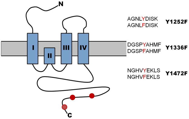
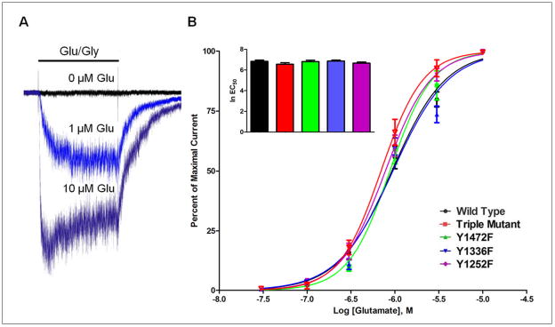
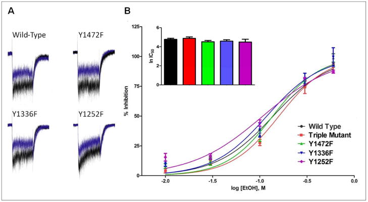
N-methyl-d-aspartate (NMDA) receptors are ion channels activated by the neurotransmitter glutamate and are highly expressed by neurons. These receptors are critical for excitatory synaptic signaling and inhibition of NMDA receptors leads to impaired cognition and learning. Ethanol inhibits NMDA currents at concentrations associated with intoxication and this action may underlie some of the behavioral effects of ethanol. Although numerous sites and mechanisms of action have been tested, the manner in which ethanol inhibits NMDA receptors remains unclear. Recent findings in the literature suggest that ethanol, via facilitation of tyrosine phosphatase activity, may dephosphorylate key tyrosine residues in the C-terminus of GluN2B subunits resulting in diminished channel function. To directly test this hypothesis, we engineered GluN2B mutants that contained phenylalanine in place of tyrosine at three different sites and transiently expressed them with the GluN1 subunit in human embryonic kidney (HEK) cells. Whole-cell patch clamp electrophysiology was used to record glutamate-activated currents in the absence and presence of ethanol (10-600 mM). All mutants were functional and did not differ from one another with respect to current amplitude, steady-state to peak ratio, or magnesium block. Analysis of ethanol dose-response curves showed no significant difference in IC50 values between wild-type receptors and Y1252F, Y1336F, Y1472F or triple Y-F mutants. These findings suggest that dephosphorylation of C-terminal tyrosine residues does not account for ethanol inhibition of GluN2B receptors.
Copyright © 2013 Elsevier Inc. All rights reserved.
Conflict of interest statement
The authors declare no competing financial interests
Figures

References
-
- Alvestad RM, Grosshans DR, Coultrap SJ, Nakazawa T, Yamamoto T, Browning MD. Tyrosine dephosphorylation and ethanol inhibition of N-methyl-D-aspartate receptor function. J Biol Chem. 2003;278(13):11020–11025. - PubMed
-
- Anders DL, Blevins T, Sutton G, Chandler LJ, Woodward JJ. Effects of c-Src tyrosine kinase on ethanol sensitivity of recombinant NMDA receptors expressed in HEK 293 cells. Alcohol Clin Exp Res. 1999;23(2):357–362. - PubMed
-
- Anders DL, Blevins T, Sutton G, Swope S, Chandler LJ, Woodward JJ. Fyn tyrosine kinase reduces the ethanol inhibition of recombinant NR1/NR2A but not NR1/NR2B NMDA receptors expressed in HEK 293 cells. J Neurochem. 1999;72(4):1389–1393. - PubMed
-
- Cavara NA, Hollmann M. Shuffling the deck anew: how NR3 tweaks NMDA receptor function. Mol Neurobiol. 2008;38(1):16–26. - PubMed
-
- Cik M, Chazot PL, Stephenson FA. Expression of NMDAR1–1a (N598Q)/NMDAR2A receptors results in decreased cell mortality. Eur J Pharmacol. 1994;266:R1–R3. - PubMed
Publication types
MeSH terms
Substances
Grants and funding
LinkOut - more resources
Full Text Sources
Other Literature Sources
Miscellaneous
