. 2013;8(1):e55035.
doi: 10.1371/journal.pone.0055035. Epub 2013 Jan 24.

PIASγ enhanced SUMO-2 modification of Nurr1 activation-function-1 domain limits Nurr1 transcriptional synergy

Affiliations

PIASγ enhanced SUMO-2 modification of Nurr1 activation-function-1 domain limits Nurr1 transcriptional synergy

Cristian Arredondo et al. PLoS One. 2013.

Abstract

Nurr1 (NR4A2) is a transcription factor that belongs to the orphan NR4A group of the nuclear receptor superfamily. Nurr1 plays key roles in the origin and maintenance of midbrain dopamine neurons, and peripheral inflammatory processes. PIASγ, a SUMO-E3 ligase, represses Nurr1 transcriptional activity. We report that Nurr1 is SUMOylated by SUMO-2 in the lysine 91 located in the transcriptional activation function 1 domain of Nurr1. Nurr1 SUMOylation by SUMO-2 is markedly facilitated by overexpressing wild type PIASγ, but not by a mutant form of PIASγ lacking its first LXXLL motif (PIASγmut1). This PIASγmut1 is also unable to interact with Nurr1 and to repress Nurr1 transcriptional activity. Interestingly, the mutant PIASγC342A that lacks SUMO ligase activity is still able to significantly repress Nurr1-dependent transcriptional activity, but not to enhance Nurr1 SUMOylation. A SUMOylation-deficient Nurr1 mutant displays higher transcriptional activity than the wild type Nurr1 only in promoters harboring more than one Nurr1 response element. Furthermore, lysine 91, the major target of Nurr1 SUMOylation is contained in a canonical synergy control motif, indicating that SUMO-2 posttranslational modification of Nurr1 regulates its transcriptional synergy in complex promoters. In conclusion, PIASγ can exert two types of negative regulations over Nurr1. On one hand, PIASγ limits Nurr1 transactivation in complex promoters by SUMOylating its lysine 91. On the other hand, PIASγ fully represses Nurr1 transactivation through a direct interaction, independently of its E3-ligase activity.


Conflict of interest statement

Competing Interests: The authors have declared that no competing interests exists.

Figures

Figure 1
Figure 1. Nurr1 is SUMOylated by SUMO-2.
(A) Schematic representation of Nurr1 (bottom) showing the position of four putative SUMOylation sites according with SUMOplotTM software analysis (middle). The table shows the sequences of the potential SUMOylation sites of rat Nurr1 sorted from the highest score. Putative SUMO acceptor lysines (K) are highlighted, and potential SUMO sites are underlined. (B) COS-7 cells were transfected with plasmids expressing HA-Nurr1, Ubc9, and SUMO-1, SUMO-2 or SUMO-3. Cells were harvested 48 hours post-transfection and lysed directly in loading buffer containing the SUMO-isopeptidase inhibitor N-ethylmaleimide 20 mM, and fractionated in SDS-PAGE. Representative western-blot with anti-HA (upper) and anti-SUMO-2 (bottom) antibodies. (C) Quantitative densitometry analysis of Nurr1-SUMO-2 signal described in (B), using Image J software. Data correspond to the mean ± S.E.M. of 3 independent experiments for each condition. Statistical significance was estimated by the non-parametric Mann-Whitney U-test. * p<0.05 (Nurr1+Ubc9+SUMO-2 v/s Nurr1). (D) Total lysates from COS-7 cells transfected with HA-Nurr1, Ubc9 and SUMO-2 were immunoprecipitated with an anti-Nurr1 antibody or control IgG. The immunoprecipitates were analyzed in western blots with anti-HA antibody. Bands for immunoglobulin are indicated as IgG heavy chain (H).
Figure 2
Figure 2. Lysine 91 is the main SUMOylation site of Nurr1.
(A) Schematic representation of full-length HA-Nurr1, and truncated isoforms HA-Nurr1ΔAF-2 (amino acids 1–353) and HA-Nurr1ΔAF-1 (amino acids 262–598). (B, C) Total extracts of COS-7 cells transfected with HA-Nurr1ΔAF-2 or HA-Nurr1ΔAF-1, plus Ubc9 and SUMO-2 were fractionated in SDS-PAGE and proteins analyzed with anti-HA (B) and anti-Nurr1 (C) antibodies. (D) Total extracts of COS-7 cells transfected with wild type HA-Nurr1 or the point mutants HA-Nurr1-K74R or HA-Nurr1-K91R were fractionated in SDS-PAGE. Western blot was performed with an anti-HA antibody.
Figure 3
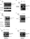
Figure 3. PIASγ enhances Nurr1 SUMOylation.
(A) Total extracts of COS-7 cells transfected with plasmids encoding HA-Nurr1, SUMO-2 and PIASγ were fractionated in SDS-PAGE and western blots were performed with anti-HA (upper), anti-SUMO-2 (middle) and anti-PIASγ (bottom) antibodies. (B) Total extracts of COS-7 cells transfected as in (A) were immunoprecipitated and immunoblotted with a monoclonal (m) anti-HA antibody. (C) Total extracts of COS-7 cells transfected with HA-Nurr1, SUMO-2, PIASγ and Flag-SENP1 or Flag-SENP1-DN were fractionated in SDS-PAGE and western blots were performed with the indicated antibodies. (D) Total extracts of COS-7 cells transfected with HA-Nurr1 or the point mutants HA-Nurr1-K74R or HA-Nurr1-K91R were fractionated in SDS-PAGE. Western blots were performed with an anti-HA antibody. (E, F) Total extracts of COS-7 cells transfected with HA-Nurr1 or HA-Nurr1-K91R plus SUMO-2 and PIASγ were immunoprecipitated with anti-HA antibody and western blot carried out with anti-Nurr1 antibody (E) or immunoprecipitated with anti-Nurr1 antibody and western blot carried out with anti-HA antibody (F). (m): monoclonal, (H): IgG heavy chain.
Figure 4
Figure 4. Point mutant PIASγC342A fails to SUMOylate Nurr1.
(A) Total extracts of COS-7 cells transfected with HA-Nurr1, SUMO-2 and PIASγ or the point mutant PIASγC342A were fractionated in SDS-PAGE and western blot assays performed with anti-HA (upper), anti-PIASγ (middle) and anti-SUMO-2 (bottom) antibodies. (B) HEK293cells were transfected with 100 ng of NBRE-3X-tk-LUC reporter and equimolar amounts of HA-Nurr1, PIASγ or PIASγC342A. Cells were harvested 48 hours post transfection and lysates assayed for luciferase activity. Results are expressed as fold of induction related to control (pcDNA3.1+) and correspond to the mean ± S.E.M. of three independent assays performed each in triplicate. Statistical significance was estimated by the non-parametric Mann-Whitney U test. *p<0.05 (Nurr1+PIASγ v/s Nurr1) and (Nurr1+ PIASγC342A v/s Nurr1). In the bottom, western blots showing the expression of recombinant proteins and actin used as loading control.
Figure 5
Figure 5. PIASγ requires the 20LXXLL24 motif to interact, SUMOylate and repress Nurr1.
(A) Schematic representation of full-length PIASγ indicating functional domains SAP: Scaffold Attachment factor-A/B acinus and PIAS; the PINIT domain; SP-(Siz/PIAS)-RING and SIM: SUMO Interaction Motif. PIASγ has two LXXLL motifs. Mutations changing leucines (L) by alanines (A) in each LXXLL domain are depicted for GST-PIASγmut1 and GST-PIASγmut2. (B) GST, GST-PIASγ, GST-PIASγmut1 and GST-PIASγmut2 retained in glutathione-agarose beads were incubated with extracts from COS-1 cells transfected with Myc-Nurr1363–598. Retained proteins were fractionated in SDS-PAGE and western blot developed with anti-Myc monoclonal antibody, revealing that only GST-PIASγ and GST-PIASγmut2 are able to interact with Nurr1. Recombinant GST-PIASγ proteins were equally loaded in each GST-pull down assays as shown by western blot using anti-PIASγ antibody (bottom). (C) Luciferase reporter assay showing that full-length PIASγmut1 lose repressor capacity over Nurr1 transactivity. HEK293 cells were transfected with the NBRE-3X-tk-LUC along with HA-Nurr1, PIASγ or PIASγmut1. After 48 hours, cells extracts were assayed for luciferase activity. Data are expressed as percentage of Nurr1 transactivation and correspond to the mean ± S.E.M of 4 independent experiments each performed in triplicates. Statistical significance was estimated by the non-parametric Mann-Whitney U test. *p<0.05 (Nurr1+PIASγ v/s Nurr1+PIASγmut1). In the bottom, western blots showing the expression of recombinant proteins and actin used as loading control. (D) Total extracts of COS-7 cells transfected with HA-Nurr1, SUMO-2 and PIASγ or PIASγmut1 were fractionated in SDS-PAGE and western blots performed with anti-HA, anti-PIASγ and actin (load control) antibodies.
Figure 6
Figure 6. SUMOylation on lysine 91 does not modify Nurr1 half-life and location.
(A) HEK293 cells were transfected with HA-Nurr1 or HA-Nurr1-K91R. Twelve hours after transfection the cells were treated with cycloheximide and harvested at the indicated hours. Total extracts were fractionated in SDS-PAGE and western blots developed with anti-HA and anti-actin (load control) antibodies. Densitometric analysis of 3 independent experiments was performed with Image J software. Data is expressed as percentage of HA-Nurr1 or HA-Nurr1-K91R expression at cero time and correspond to the mean ± S.E.M. (B) Representative western blots of HA-Nurr1 and HA-Nurr1-K91R during cycloheximide treatment. (C) PC12 cells were transfected with the indicated plasmids. Fixed cells were subjected to double immunofluorescence protocols using HA and PIASγ antibodies. Alexa 594 (red) second antibody was used to visualize HA and Alexa 488 (green) second antibody was used to visualize PIASγ. Cells were examined by deconvolution microscopy. (D) Colocalization of Nurr1 or the mutant Nurr1-K91R with PIASγ using van-Steensel analysis .
Figure 7
Figure 7. Lysine 91 of Nurr1 is in a synergy control (SC) motif.
(A) Consensus SC motif and putative Nurr1 SC motif. (B) HEK293 cells were transfected with 100 ng of 1NBRE-Luc (one NBRE element), 3NBRE-Luc (three NBRE elements) or NBRE-3X-tk-LUC (three NBRE elements) reporters and equimolar amounts of HA-Nurr1 or point mutant HA-Nurr1-K91R. Cells were harvested 48 hours post-transfection and lysates assayed for luciferase activity. Results are expressed as fold of induction related to control (pCGN, empty vector) and correspond to the mean ± S.E.M. of three independent assays performed each in triplicate. Scale in left axis is for 1NBRE-Luc and 3NBRE-Luc reporters' activity, and scale in right axis is for NBRE-3X-tk-LUC reporter activity. Statistical significance was estimated by the non-parametric Mann-Whitney U test *p<0.05 (HA-Nurr1-K91R v/s HA-Nurr1). (C) HEK293cells were transfected with 100 ng of 1NBRE-Luc or 3NBRE-Luc reporters and equimolar amounts of HA-Nurr1ΔAF-2 or point mutant HA-Nurr1ΔAF-2-K91R. Cells were harvested 48 hours post-transfection and lysate assayed for luciferase activity. Results are expressed as fold of induction related to control (pCGN) and correspond to the mean ± S.E.M. of three independent assays performed each in triplicate.

Similar articles

Cited by

References

    1. Perlmann T, Wallén-Mackenzie A (2004) Nurr1, an orphan nuclear receptor with essential functions in developing dopamine cells. Cell Tissue Res 318: 45–52. - PubMed
    1. Maxwell MA, Muscat GE (2006) The NR4A subgroup: immediate early response genes with pleiotropic physiological roles. Nucl Recept Signal 4: e002. - PMC - PubMed
    1. Maruyama K, Tsukada T, Ohkura N, Bandoh S, Hosono T, et al. (1998) The NGFI-B subfamily of the nuclear receptor superfamily. Int J Oncol 12: 1237–1243. - PubMed
    1. Wang Z, Benoit G, Liu J, Prasad S, Aarnisalo P, et al. (2003) Structure and function of Nurr1 identifies a class of ligand-independent nuclear receptors. Nature 423: 555–560. - PubMed
    1. Castro DS, Arvidsson M, Bondesson-Bolin M, Perlmann T (1999) Activity of the Nurr1 carboxy-terminal domain depends on cell type and integrity of the activation function 2. J Biol Chem 274: 37483–37490. - PubMed

Publication types

MeSH terms

Substances

LinkOut - more resources