. 2013 Apr;21(4):860-7.
doi: 10.1038/mt.2013.9. Epub 2013 Jan 29.

The use of scaffold-free cell sheet technique to refine mesenchymal stromal cell-based therapy for heart failure

Affiliations

The use of scaffold-free cell sheet technique to refine mesenchymal stromal cell-based therapy for heart failure

Takuya Narita et al. Mol Ther. 2013 Apr.

Abstract

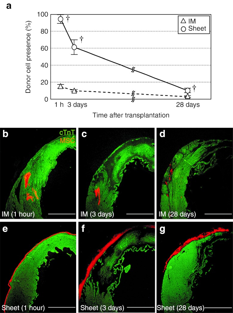
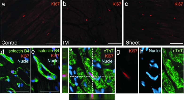
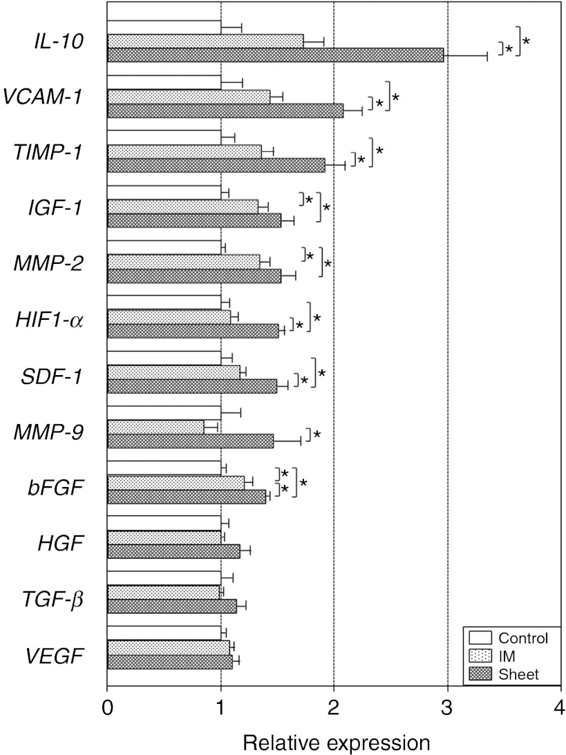
Transplantation of bone marrow-derived mesenchymal stromal cells (MSCs) is an emerging treatment for heart failure based on their secretion-mediated "paracrine effects". Feasibility of the scaffoldless cell sheet technique to enhance the outcome of cell transplantation has been reported using other cell types, though the mechanism underpinning the enhancement remains uncertain. We here investigated the role of this innovative technique to amplify the effects of MSC transplantation with a focus on the underlying factors. After coronary artery ligation in rats, syngeneic MSCs were grafted by either epicardial placement of MSC sheets generated using temperature-responsive dishes or intramyocardial (IM) injection. Markedly increased initial retention boosted the presence of donor MSCs persistently after MSC sheet placement although the donor survival was not improved. Most of the MSCs grafted by the cell sheet technique remained resided on the epicardial surface, but the epicardium quickly regressed and new vessels sprouted into the sheets, assuring the permeation of paracrine mediators from MSCs into the host myocardium. In fact, there was augmented upregulation of various paracrine effect-related genes and signaling pathways in the early phase after MSC sheet therapy. Correspondingly, more extensive paracrine effects and resultant cardiac function recovery were achieved by MSC sheet therapy. Further development of this approach towards clinical application is encouraged.


Figures

Figure 1
Figure 1
Improved donor cell retention and distribution by MSC sheet therapy. (a) Quantitative assessments by using the detection of the male-specific sry gene in the female heart showed that the initial retention (at 1 hour) and successive presence of donor cells (% of the total donor cell number grafted) were improved in the Sheet group compared with the IM group. P < 0.05 versus the IM group, mean ± SEM for n = 4–6 in each point. Histological evaluation showed that donor cell distribution was distinct between groups; (bd) intramyocardial cell cluster formation in the IM group (for 1 hour, days 3 and 28, respectively) versus (eg) retention on the epicardial surface in the Sheet group (for 1 hour, days 3 and 28, respectively). Orange signal for MSCs (DiI); green for cTnT. Bar = 1 mm. cTnT, cardiac troponin-T; IM, intramyocardial injection; MSC, mesenchymal stromal cell.
Figure 2
Figure 2
Donor cell apoptosis after MSC sheet therapy. (a) Immunolabeling for cleaved caspase-3 detected a similar frequency of apoptotic donor cells in both treatment groups at day 3. (b) A representative image of apoptotic donor cells (yellow arrow) after MSC sheet therapy is shown. Enlarged images of white-gated area in b are presented in cf with each marker separate and merged. Green signal for cleaved caspase-3; orange for MSCs (DiI); blue for nuclei (DAPI). Bar = 30 µm in b. DAPI, 4′,6-diamidino-2-phenylindole; IM, intramyocardial injection; MSC, mesenchymal stromal cell.
Figure 3
Figure 3
Donor cell behaviors and changes in the epicardium after MSC sheet therapy. (ad) Isolectin B4 staining demonstrated that the vasculature in MSC sheets contained both host-derived (DiI) and donor MSC-derived (DiI+) endothelial cells at day 28 after transplantation of DiI+ sheets. (e) ICAM1 staining identified the epicardium in the Control group, (f) though this was absent by day 3 after MSC sheet therapy. (g) CD31 staining detected host-derived (Dil) endothelium sprouting into the MSC sheets at day 3. (h) The majority of donor MSCs retained on the surface of the heart (as in f), but a small number of MSCs had migrated into the host myocardium at day 3. Green signal for isolectin B4 in a,b, ICAM-1 in e,f or CD31 in g; orange for MSCs (DiI); blue for nuclei (DAPI). Bar = 30 µm in ad, 50 µm in eh. DAPI, 4′,6-diamidino-2-phenylindole; ICAM, intercellular adhesion molecule; MSC, mesenchymal stromal cell.
Figure 4
Figure 4
Histological recovery of post-MI failing cardiac tissues by MSC sheet therapy. (a) Histological studies detected reduced infarct size (Supplementary Figure S5a–c), (b) increased capillary density (Supplementary Figure S5d–f), (c) reduced cardiomyocyte hypertrophy (Supplementary Figure S5g–i), and (d) attenuated collagen deposition (Supplementary Figure S5j–l) at day 28 in the Sheet group compared with the Control group (and to the IM group in most points).*P < 0.05, mean ± SEM for n = 4–5 in each point. IM, intramyocardial injection; MI, myocardial infarction; MSC, mesenchymal stromal cell.
Figure 5
Figure 5
Improved endogenous regenerative activity by MSC sheet therapy. The number of Ki67+ cells was increased in the Sheet group compared with the IM and Control groups. Representative images of Ki67+ cells in the border areas of the (a) Control, (b) IM, and (c) Sheet groups at day 28 are shown (Table 2). Ki67+/isolectin B4+ cells were found in (d) capillaries as well as in (e) larger vessels after MSC sheet therapy. There were an increased number of Ki67+/cTnT+ proliferating cardiomyocytes in the Sheet group. (fi) A representative image of Ki67+/cTnT+ cells after MSC sheet therapy is presented for each marker and merged. Green signal for isolectin B4 in d,e, cTnT in f,i; orange for Ki67; blue for nuclei (DAPI). Bar =100 µm in ac and 30 µm in df. cTnT, cardiac troponin-T; DAPI, 4′,6-diamidino-2-phenylindole; IM, intramyocardial injection; MSC, mesenchymal stromal cell.
Figure 6
Figure 6
Amplified upregulation of paracrine effect-relevant genes by MSC sheet therapy. Quantitative RT-PCR screening detected upregulation of a group of genes likely to be relevant to the MSC-derived paracrine effects in the Sheet group, and in the IM group to a lesser extent, at day 3. All expression levels were normalized to that in the Control group, which was assigned a value of 1.0. *P < 0.05, mean ± SEM for n = 5–6 in each group. IM, intramyocardial injection; MSC, mesenchymal stromal cell; RT-PCR, reverse transcription-PCR.

References

    1. Menasche P. Cardiac cell therapy: lessons from clinical trials. J Mol Cell Cardiol. 2011;50:258–265. - PubMed
    1. Passier R, van Laake LW., and, Mummery CL. Stem-cell-based therapy and lessons from the heart. Nature. 2008;453:322–329. - PubMed
    1. Choi YH, Kurtz A., and, Stamm C. Mesenchymal stem cells for cardiac cell therapy. Hum Gene Ther. 2011;22:3–17. - PubMed
    1. Boyle AJ, McNiece IK., and, Hare JM. Mesenchymal stem cell therapy for cardiac repair. Methods Mol Biol. 2010;660:65–84. - PubMed
    1. Gnecchi M, He H, Liang OD, Melo LG, Morello F, Mu H.et al. (2005Paracrine action accounts for marked protection of ischemic heart by Akt-modified mesenchymal stem cells Nat Med 11367–368. - PubMed

Publication types