. 2013 Feb 4;200(3):259-70.
doi: 10.1083/jcb.201211017. Epub 2013 Jan 28.

Structural basis of tubulin tyrosination by tubulin tyrosine ligase

Affiliations

Structural basis of tubulin tyrosination by tubulin tyrosine ligase

Andrea E Prota et al. J Cell Biol. .

Abstract

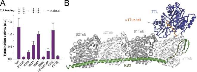
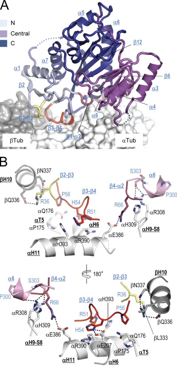
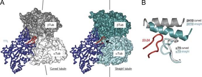
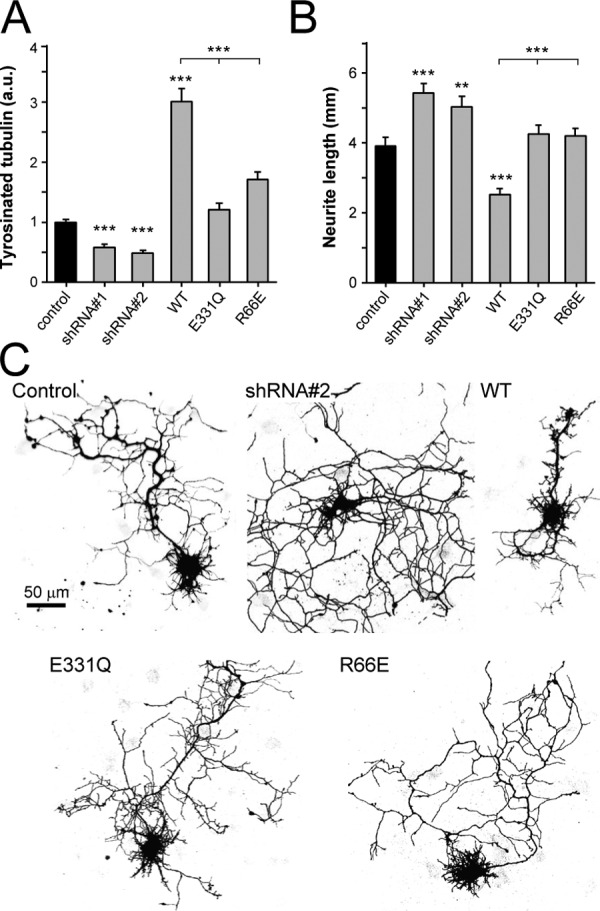
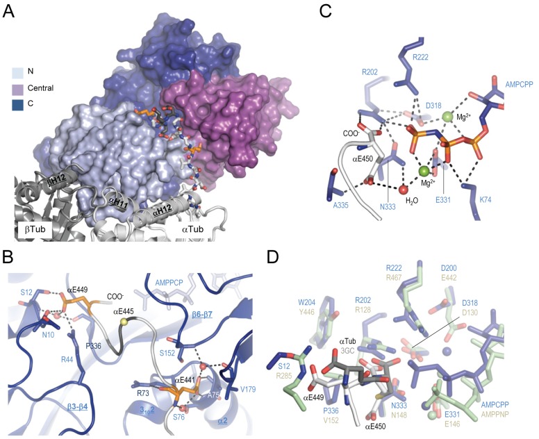
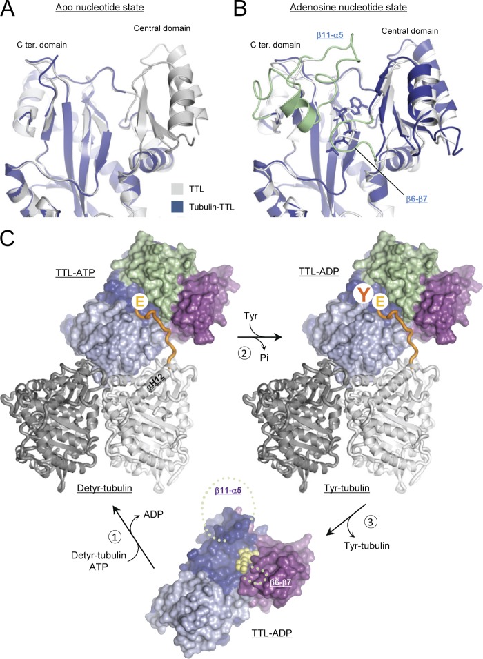
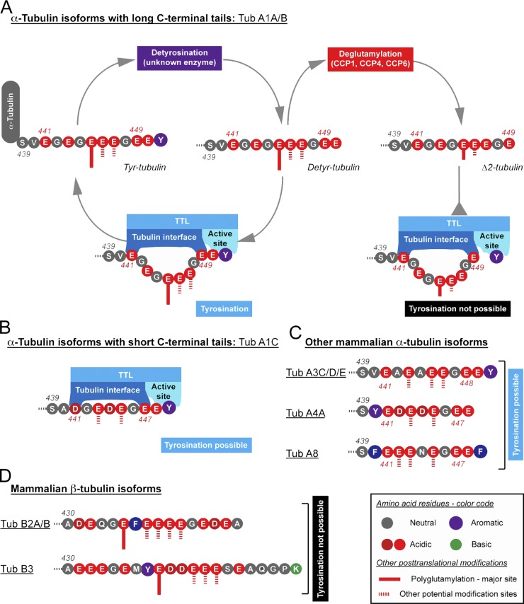
Tubulin tyrosine ligase (TTL) catalyzes the post-translational retyrosination of detyrosinated α-tubulin. Despite the indispensable role of TTL in cell and organism development, its molecular mechanism of action is poorly understood. By solving crystal structures of TTL in complex with tubulin, we here demonstrate that TTL binds to the α and β subunits of tubulin and recognizes the curved conformation of the dimer. Biochemical and cellular assays revealed that specific tubulin dimer recognition controls the activity of the enzyme, and as a consequence, neuronal development. The TTL-tubulin structure further illustrates how the enzyme binds the functionally crucial C-terminal tail sequence of α-tubulin and how this interaction catalyzes the tyrosination reaction. It also reveals how TTL discriminates between α- and β-tubulin, and between different post-translationally modified forms of α-tubulin. Together, our data suggest that TTL has specifically evolved to recognize and modify tubulin, thus highlighting a fundamental role of the evolutionary conserved tubulin tyrosination cycle in regulating the microtubule cytoskeleton.


Figures

Figure 1.
Figure 1.
Tubulin binding, enzymatic activity, and overall T2R–TTL structure. (A) Enzyme activity tests (bars) and T2R-binding properties (symbols on top of the bars) of wild-type (WT) and different mutants of TTL. Error bars indicate SEM. The T2R-binding properties were assessed by size exclusion chromatography (see Fig. S1 A for representative data). The binding strength of TTL variants are classified from weak binding (one cross) to strong binding (four crosses). Minus sign, no binding; n.d., not determined. (B) Overall view of the T2R–TTL complex structure in ribbon representation. α-Tubulin, β-tubulin, TTL, and the stathmin-like domain of RB3 are shown in light gray, dark gray, blue, and green, respectively. The C-terminal tail region of α1-tubulin that is bound to TTL is highlighted in orange.
Figure 2.
Figure 2.
Tubulin recognition by TTL. (A) Global view of the tubulin–TTL interaction. The N-terminal (residues 1–71), central (residues 72–188), and C-terminal (residues 189–378) domains of TTL (ribbon representation) are colored in light blue, magenta, and deep blue, respectively. The interacting TTL loops β2- β3, β3- β4, and β4-α2, and the N-terminal part of helix α6 are highlighted in pale yellow, red, raspberry, and pink, respectively. The αβ-tubulin heterodimer is shown in surface representation and selected TTL elements are labeled. For simplicity, the C-terminal tail of α-tubulin is not shown. (B) Two close-up views 180° apart of the tubulin–TTL interface shown in A in ribbon representation. Interacting residues are shown in stick representation and are labeled in black (tubulin) and blue (TTL). Selected secondary structure elements of tubulin and TTL are labeled in bold black and blue underlined letters, respectively. The loops of TTL that bind tubulin are colored as in A. Oxygen and nitrogen atoms are colored in red and blue, respectively, carbon atoms in the color of the secondary structure elements depicted. Hydrogen bonds are depicted as black dashed lines. For simplicity, the Arg46-αGlu433 TTL–α-tubulin salt bridge and the interfacial water–mediated hydrogen bonding network is not depicted.
Figure 3.
Figure 3.
TTL recognizes the curved conformation of αβ-tubulin. (A) TTL (blue) bound to the curved (left) and straight (right) conformation of αβ-tubulin. α- and β-tubulin are shown in light and dark gray, and cyan and deep teal for the curved and straight conformations, respectively. The arrow in the right panel points to the site in which the β3-β4 loop of TTL (red) clashes into the straight conformation of tubulin. The straight tubulin structure corresponds to Protein Data Bank accession no. 1JFF (Löwe et al., 2001). (B) Superimposition of the TTL-bound curved tubulin conformation (T2R–TTL) onto the straight tubulin structure. The N-terminal nucleotide-binding and the C-terminal domains of α-tubulin were used for the superimposition (Ravelli et al., 2004). For simplicity only the T5 loop of α-tubulin (light gray and cyan for the curved and straight tubulin conformations, respectively), the H10 helix of β-tubulin (dark gray and deep teal for the curved and straight tubulin conformations, respectively), and the β3-β4 loop of TTL (red) are shown.
Figure 4.
Figure 4.
Functional analysis of TTL in hippocampal neurons. (A) Quantification of tyrosinated tubulin staining intensities in neurons transfected with GFP (control), TTL shRNA, wild-type, or mutant forms of TTL. The average intensity of the staining in the cell body was measured and the ratio of the signal in transfected versus neighboring control neurons was calculated for each image. 15–25 cells were analyzed for each condition. Error bars indicate SEM; ***, P < 0.0005. (B) Quantification of total neurite length of neurons transfected with control, TTL shRNA, wild-type, or mutant forms of TTL. 15–30 cells were analyzed for each condition. Error bars indicate SEM; *, P < 0.05; ***, P < 0.0005. (C) Representative images of hippocampal neurons (DIV5) cotransfected with β-galactosidase (to visualize morphology) and control, TTL shRNA, wild-type, or mutant forms of TTL. Bar, 50 µm.
Figure 5.
Figure 5.
Recognition of the C-terminal tail of α-tubulin by TTL. (A) Overview of the interaction between the α-tubulin tail (sticks) and TTL (surface). The two α-tubulin residues αGlu441 and αGlu449, which anchor the tail sequence of α-tubulin on TTL, are highlighted in orange. (B) Close-up view of the interaction between TTL (blue ribbon) and the C-terminal tail of α-tubulin (gray ribbon). Interacting residues are shown in stick representation. αGlu441 and αGlu449 are highlighted in orange. The position of αGlu445, the major modification site of α-tubulin, is highlighted by a yellow sphere. Three key water molecules are shown as red spheres. Note that αGlu445, αGlu446, and αGlu447 are not well defined in the electron density (see Fig. S4 A) and are thus modeled and shown in dark gray in A and B. (C) Close-up view of the active site of TTL in the T2R–TTL complex in stick representation. A water molecule and two magnesium ions are depicted as red and green spheres, respectively. (D) Superimposition of the active site of TTL (blue) bound to the nonhydrolyzable ATP analogue AMPPCP and the tail of α-tubulin (light gray) onto the one of glutathione synthase (green; Protein Data Bank accession no. 1M0W) bound to the nonhydrolyzable ATP analogue AMPPNP and gamma-glutamylcysteine (3GC; dark gray). Key residues are shown in stick representation. For simplicity only residue side chains are shown. Spheres depict magnesium ions. See Fig. 2 for additional information on labels, symbols, and color code.
Figure 6.
Figure 6.
Molecular mechanism of tubulin tyrosination by TTL. (A and B) Superimposition of TTL in the tubulin-bound (blue) and -unbound (gray; Protein Data Bank accession nos. 3TIG and 3TII, respectively) states in cartoon representations, and in the absence (A) or presence (B) of nonhydrolyzable ATP analogues. In B, the β11-α5 loop and a large portion of the central domain of TTL, structural elements that get structured upon binding of both tubulin and adenosine nucleotide, are highlighted in pale green. (C) Enzymatic cycle of TTL. (1) Binding of TTL-ADP (ground state) to detyrosinated tubulin (Detyr-tubulin) and exchanging ADP (in yellow sphere representation) by ATP induces the structuring of the β6-β7 and β11-α5 loops of TTL (indicated by pale green dashed lines). As a result, the ATP molecule gets buried and an extended cavity is formed, which specifically recognizes the incoming C-terminal tail of α-tubulin (highlighted in orange). (2) In the presence of tyrosine and upon ATP hydrolysis, tyrosinated tubulin (Tyr-tubulin), ADP, and inorganic phosphate (Pi) are produced. (3) Release of Tyr-tubulin from TTL restores the ground state of TTL-ADP. This could be achieved by phosphorylation or by the intrinsic thermodynamic properties of the moderately stable tubulin–TTL complex (Szyk et al., 2011), which suggests a fast dissociation rate. The αβ-tubulin heterodimer (α- and β-tubulin in light and dark gray, respectively) is shown in cartoon representation; TTL is shown in surface representation. TTL domains are colored as in Fig. 2 A.
Figure 7.
Figure 7.
Schematic representation of the interplay between different post-translational modifications and isoforms of tubulin, and the involvement of TTL. (A) α-Tubulin is detyrosinated by a yet unknown enzyme and gives rise to detyrosinated tubulin (Detyr-tubulin). The C-terminal tail of detyrosinated tubulin originating from the most prominent α-tubulin isoforms Tub A1A or Tub A1B is anchored by TTL via the acidic residues αGlu441 and αGlu449. The main site of polyglutamylation, αGlu445, is localized between these two residues and therefore does not interfere with the formation of the tubulin–TTL complex. Further transformation of detyrosinated tubulin into Δ2-tubulin by deglutamylases of the carboxy peptidase (CCP) family does not interfere with the binding of tubulin to TTL, but the tyrosination reaction is not anymore possible as the C terminus of Δ2-tubulin (αGlu449 in Tub A1A and Tub A1B) is too distant from the binding site of tyrosine. (B) A shorter isoform of α-tubulin, Tub A1C, can still bind TTL as the two binding sites on the enzyme are close enough. As in Tub A1A and A1B (A), the potential sites of polyglutamylation (which are most likely also the sites of polyglycylation) are localized in regions that are not involved in the formation of the tubulin–TTL complex. (C) Depiction of C-terminal tails of remaining mammalian α-tubulin isoforms. All these isoforms possess the two acidic “anchoring” residues necessary for TTL binding, and can therefore potentially be tyrosinated. Tub A4A is gene encoded in the detyrosinated form. Tub A8 carries a C-terminal phenylalanine instead of a tyrosine. To act as a substrate for TTL, it is necessary that phenylalanine can be enzymatically removed, which is currently not known. (D) Depiction of C-terminal tails of two major brain β-tubulin isoforms. Note that none of the β-tubulin isoforms contain the C-terminal Glu-Glu dipeptide sequence necessary for tyrosination by TTL.

References

    1. Adams P.D., Afonine P.V., Bunkóczi G., Chen V.B., Davis I.W., Echols N., Headd J.J., Hung L.W., Kapral G.J., Grosse-Kunstleve R.W., et al. 2010. PHENIX: a comprehensive Python-based system for macromolecular structure solution. Acta Crystallogr. D Biol. Crystallogr. 66:213–221 10.1107/S0907444909052925 - DOI - PMC - PubMed
    1. Alexander J.E., Hunt D.F., Lee M.K., Shabanowitz J., Michel H., Berlin S.C., MacDonald T.L., Sundberg R.J., Rebhun L.I., Frankfurter A. 1991. Characterization of posttranslational modifications in neuron-specific class III beta-tubulin by mass spectrometry. Proc. Natl. Acad. Sci. USA. 88:4685–4689 10.1073/pnas.88.11.4685 - DOI - PMC - PubMed
    1. Andreu J.M. 2007. Large scale purification of brain tubulin with the modified Weisenberg procedure. Methods Mol. Med. 137:17–28 10.1007/978-1-59745-442-1_2 - DOI - PubMed
    1. Arce C.A., Rodriguez J.A., Barra H.S., Caputo R. 1975. Incorporation of L-tyrosine, L-phenylalanine and L-3,4-dihydroxyphenylalanine as single units into rat brain tubulin. Eur. J. Biochem. 59:145–149 10.1111/j.1432-1033.1975.tb02435.x - DOI - PubMed
    1. Badin-Larçon A.C., Boscheron C., Soleilhac J.M., Piel M., Mann C., Denarier E., Fourest-Lieuvin A., Lafanechère L., Bornens M., Job D. 2004. Suppression of nuclear oscillations in Saccharomyces cerevisiae expressing Glu tubulin. Proc. Natl. Acad. Sci. USA. 101:5577–5582 10.1073/pnas.0307917101 - DOI - PMC - PubMed

Publication types

Associated data