. 2013 Jan;9(1):e1003141.
doi: 10.1371/journal.ppat.1003141. Epub 2013 Jan 24.

Point mutations in FimH adhesin of Crohn's disease-associated adherent-invasive Escherichia coli enhance intestinal inflammatory response

Affiliations

Point mutations in FimH adhesin of Crohn's disease-associated adherent-invasive Escherichia coli enhance intestinal inflammatory response

Nicolas Dreux et al. PLoS Pathog. 2013 Jan.

Abstract

Adherent-invasive Escherichia coli (AIEC) are abnormally predominant on Crohn's disease (CD) ileal mucosa. AIEC reference strain LF82 adheres to ileal enterocytes via the common type 1 pili adhesin FimH and recognizes CEACAM6 receptors abnormally expressed on CD ileal epithelial cells. The fimH genes of 45 AIEC and 47 non-AIEC strains were sequenced. The phylogenetic tree based on fimH DNA sequences indicated that AIEC strains predominantly express FimH with amino acid mutations of a recent evolutionary origin - a typical signature of pathoadaptive changes of bacterial pathogens. Point mutations in FimH, some of a unique AIEC-associated nature, confer AIEC bacteria a significantly higher ability to adhere to CEACAM-expressing T84 intestinal epithelial cells. Moreover, in the LF82 strain, the replacement of fimH(LF82) (expressing FimH with an AIEC-associated mutation) with fimH(K12) (expressing FimH of commensal E. coli K12) decreased the ability of bacteria to persist and to induce severe colitis and gut inflammation in infected CEABAC10 transgenic mice expressing human CEACAM receptors. Our results highlight a mechanism of AIEC virulence evolution that involves selection of amino acid mutations in the common bacterial traits, such as FimH protein, and leads to the development of chronic inflammatory bowel disease (IBD) in a genetically susceptible host. The analysis of fimH SNPs may be a useful method to predict the potential virulence of E. coli isolated from IBD patients for diagnostic or epidemiological studies and to identify new strategies for therapeutic intervention to block the interaction between AIEC and gut mucosa in the early stages of IBD.


Conflict of interest statement

The authors have declared that no competing interests exist.

Figures
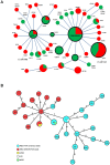
Figure 1
Figure 1. FimH protein Zonal Phylogeny and MLST tree of AIEC used in this study.
(A) Each circle represents a FimH variant coded by phylogenetically distinct fimH allele(s). Circle size reflects the number of strains (from 1 to 12) carrying the corresponding FimH variant. Consensus, the most common and evolutionarily primary FimH variant. All amino acid changes indicated are derivatives from the consensus variant. Circles inside the black rings represent evolutionarily fixed FimH variants coded by multiple phylogenetically linked fimH alleles with silent variations only. The rest are evolutionarily recent FimH variants coded by a single fimH allele. In red – the proportion of AIEC strains carrying the corresponding FimH variant. In green – the proportion of non-AIEC strains. (B) Minimum spanning tree based on the MLST allelic profiles portraying the clonal distribution of 45 AIEC strains and 3 reference strains. Each dot represents a given sequence type (ST) and the size of the circle is proportional to the number of strains analyzed. Connecting lines of increasing length and the numbers on these lines demonstrate the number of different alleles between two STs. The color of the dots represents the different AIEC clades and the reference strains.
Figure 2
Figure 2. Adhesion ability of AIEC and non-AIEC strains with regard to fimH clade and CEACAM6 expression.
(A) Cell-associated bacteria were quantified using non differentiated T84 cells after a 3 H infection period, and results were analyzed with regard to fimH clade. (B) Cell-associated AIEC bacteria belonging to the fimH S70/N78 clade were quantified using undifferentiated and differentiated T84 cells after a 3 H infection period. (C) Western blot analysis of whole protein extracts from non-differentiated and differentiated T84 cells using anti-CEACAM6 and anti-GAPDH antibodies. (D) Cell-associated bacteria (AIEC and non-AIEC bacteria belonging to the fimH S70/N78 clade) were quantified using differentiated T84 cells after a 3 H infection period. (E) Yeast agglutination titer of AIEC and non-AIEC strains belonging to the fimH S70/N78 clade. (F) Transmission electron micrograph of negatively stained AIEC and non-AIEC bacteria belonging to the fimH S70/N78 clade, magnification ×25 000.
Figure 3
Figure 3. Construction of fimH chromosomal mutants.
(A) Construction of fimH chromosomal mutants. (B) Electron microscopy examination of AIEC LF82 bacteria, LF82-ΔfimH isogenic mutant, LF82-ΔfimH/fimHLF82, LF82-ΔfimH/fimHK12, LF82-ΔfimH/fimH7082, LF82-ΔfimH/fimHLF28, LF82-ΔfimH/fimHLF16, LF82-ΔfimH/fimH7049, LF82-ΔfimH/fimHLF31, LF82-ΔfimH/fimH7136; LF82-ΔfimH/fimHLF73 showing flagella and type 1 pili expression (magnification ×40 000). (C) Yeast agglutination titer of AIEC LF82 bacteria, LF82-Δ fimH isogenic mutant and fimH chromosomal mutants.
Figure 4
Figure 4. Impact of FimH amino acid substitutions on AIEC adhesion ability.
Cell-associated bacteria were quantified using undifferentiated (A) or differentiated (B) T84 cells after a 3 H infection period. WT represents the original AIEC LF82 reference strain and ΔfimH represents the LF82-ΔfimH isogenic mutant. The other constructs were made in the LF82-ΔfimH isogenic mutant, in which various fimH variants were reintroduced at the fimH locus. Each value is the mean ± SEM of at least four separate experiments (* P<0.05; ** P<0.001; *** P<0.001). (C) Relative ability to bind Man1 (1 M/3 M binding ratio) with regard to undifferentiated T84 index adhesion of the various fimH chromosomal mutants constructed. (D) Bacterial binding to 1M-BSA under various shear stresses (pN/m cm2) was evaluated by measuring bacterial accumulation over 3 min.
Figure 5
Figure 5. Bacterial colonization, colonic mucosa inflammation and translocation in CEABAC10 mice infected with LF82ΔfimH/fimHLF82 or LF82ΔfimH/fimHK12 mutants.
(A) Quantification of LF82-ΔfimH/fimHLF82 (black square) or LF82-ΔfimH/fimHK12 (white square) bacteria in the feces of CEABAC10 mice receiving 0.25% DSS in drinking water after oral infection with 109 bacteria on day 0. (B) Quantification of colonic mucosal-associated LF82-ΔfimH/fimHLF82 or LF82-ΔfimH/fimHK12 bacteria on day of sacrifice. (C) Quantification of bacteria on day of sacrifice in the liver and spleen. (D) DAI was performed for CEABAC10 transgenic mice infected with (black square) LF82ΔfimH/fimHLF82 or with (white square) LF82ΔfimH/fimHK12. (E) Il-1β secretion by colonic mucosa. (F) Histopathological scoring for several parameters of colonic inflammation was performed for CEABAC10 transgenic mice infected with (black square) LF82ΔfimH/fimHLF82 or with (white square) LF82ΔfimH/fimHK12. * P<0.05; ** P<0.01; and *** P<0.001.
Figure 6
Figure 6. Bacterial persistence and IL-1β secretion by colonic mucosa of CEABAC10 mice according to FimH sequence.
(A) Quantification of LF82-ΔfimH/fimHLF82 (black square), LF82-ΔfimH/fimH7082 (white circle), LF82-ΔfimH/fimHK12 (white square) and LF82-ΔfimH/fimHLF28 (white triangle) bacteria in the feces of CEABAC10 mice infected with 109 bacteria on day 3 post-infection. (B) Quantification of IL-1β release by colonic loops infected with LF82 bacteria expressing fimH from LF82 or 7082 (S70/N78 clade) and from K12 or LF28 (consensus clade). * P<0.05; ** P<0.01.
Figure 7
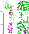
Figure 7. Positions 73, 158 and 166 in the crystal structure of FimH in complex with FimG (pdb id: 3JWN).
Carbon atoms are shown in green for FimH and in violet for FimG. Oxygen atoms are shown in red and nitrogen atoms in blue. The hydrogen bonds are shown in red dashed lines, and close contact in blue dashed lines.

References

    1. Kaser A, Zeissig S, Blumberg RS (2010) Inflammatory bowel disease. Annu Rev Immunol 28: 573–621. - PMC - PubMed
    1. Xavier RJ, Podolsky DK (2007) Unravelling the pathogenesis of inflammatory bowel disease. Nature 448: 427–434. - PubMed
    1. Darfeuille-Michaud A, Boudeau J, Bulois P, Neut C, Glasser AL, et al. (2004) High prevalence of adherent-invasive Escherichia coli associated with ileal mucosa in Crohn's disease. Gastroenterology 127: 412–421. - PubMed
    1. Darfeuille-Michaud A, Neut C, Barnich N, Lederman E, Di Martino P, et al. (1998) Presence of adherent Escherichia coli strains in ileal mucosa of patients with Crohn's disease. Gastroenterology 115: 1405–1413. - PubMed
    1. Eaves-Pyles T, Allen CA, Taormina J, Swidsinski A, Tutt CB, et al. (2008) Escherichia coli isolated from a Crohn's disease patient adheres, invades, and induces inflammatory responses in polarized intestinal epithelial cells. Int J Med Microbiol 298: 397–409. - PubMed

Publication types

MeSH terms