

Pathogenesis of Modoc virus (Flaviviridae; Flavivirus) in persistently infected hamsters
- PMID: 23358636
- PMCID: PMC3592524
- DOI: 10.4269/ajtmh.12-0110
Pathogenesis of Modoc virus (Flaviviridae; Flavivirus) in persistently infected hamsters
Abstract
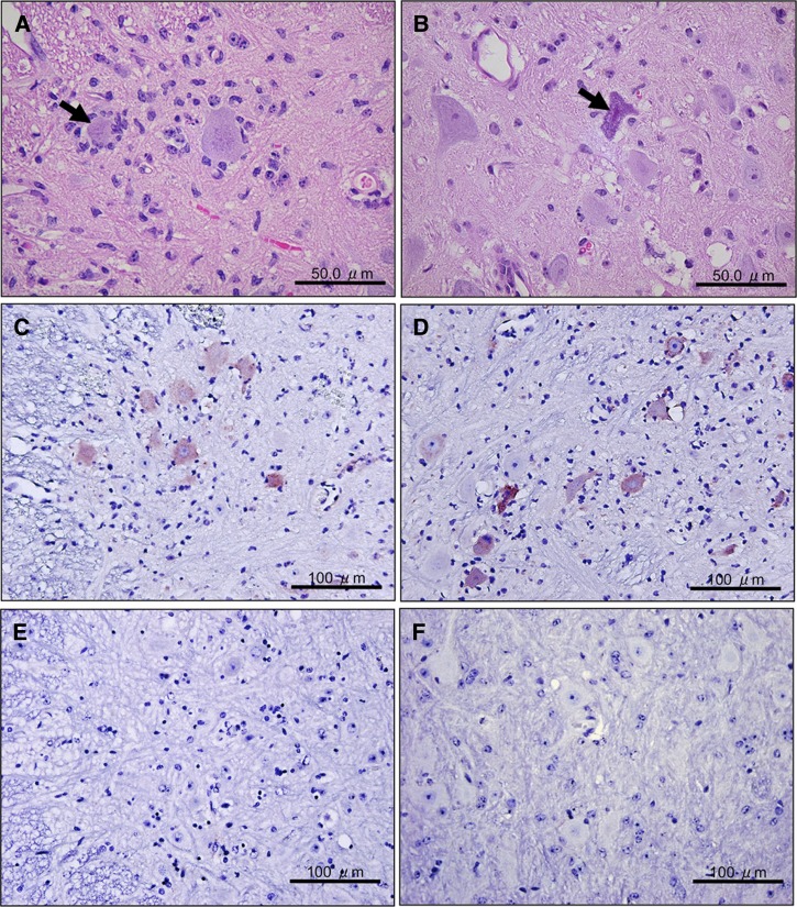
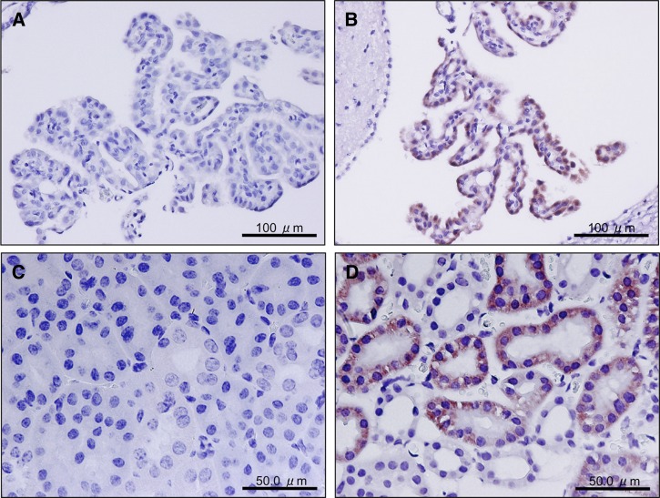
The long-term persistence of Modoc virus (MODV) infection was investigated in a hamster model. Golden hamsters (Mesocricetus auratus) were infected by subcutaneous inoculation with MODV, in which fatal encephalitis developed in 12.5% (2 of 16). Surviving hamsters shed infectious MODV in their urine during the first five months after infection, and infectious MODV was recovered by co-cultivation of kidney tissue up to eight months after infection. There were no histopathologic changes observed in the kidneys despite detection of viral antigen for 250 days after infection. Mild inflammation and neuronal degeneration in the central nervous system were the primary lesions observed during early infection. These findings confirm previous reports of persistent flavivirus infection in animals and suggest a mechanism for the maintenance of MODV in nature.
Figures
References
-
- Johnson HN. Ecological implications of antigenically related mammalian viruses for which arthropod vectors are unknown and avian-associated soft tick viruses. Jpn J Med Sci Biol. 1967;20:160–166. - PubMed
-
- Karabatsos N. International Catalogue of Arboviruses Including Certain Other Viruses of Vertebrates. Third edition. San Antonio, TX: American Society of Tropical Medicine and Hygiene; 1984. pp. 691–692. - PubMed
-
- Zarnke RL, Yuill TM. Modoc-like virus isolated from wild deer mice (Peromyscus maniculatus) J Wildl Dis. 1985;21:94–99. - PubMed
-
- Fairbrother A, Yuill TM. Experimental infection and horizontal transmission of Modoc virus in deer mice (Peromyscus maniculatus) J Wildl Dis. 1987;23:179–185. - PubMed
Publication types
MeSH terms
Substances
Grants and funding
LinkOut - more resources
Full Text Sources
Other Literature Sources
