Comparative Study
. 2013 Apr 1;73(7):2368-78.
doi: 10.1158/0008-5472.CAN-12-3424. Epub 2013 Jan 28.

ST6Gal-I protein expression is upregulated in human epithelial tumors and correlates with stem cell markers in normal tissues and colon cancer cell lines

Affiliations
Comparative Study

ST6Gal-I protein expression is upregulated in human epithelial tumors and correlates with stem cell markers in normal tissues and colon cancer cell lines

Amanda F Swindall et al. Cancer Res. .

Abstract

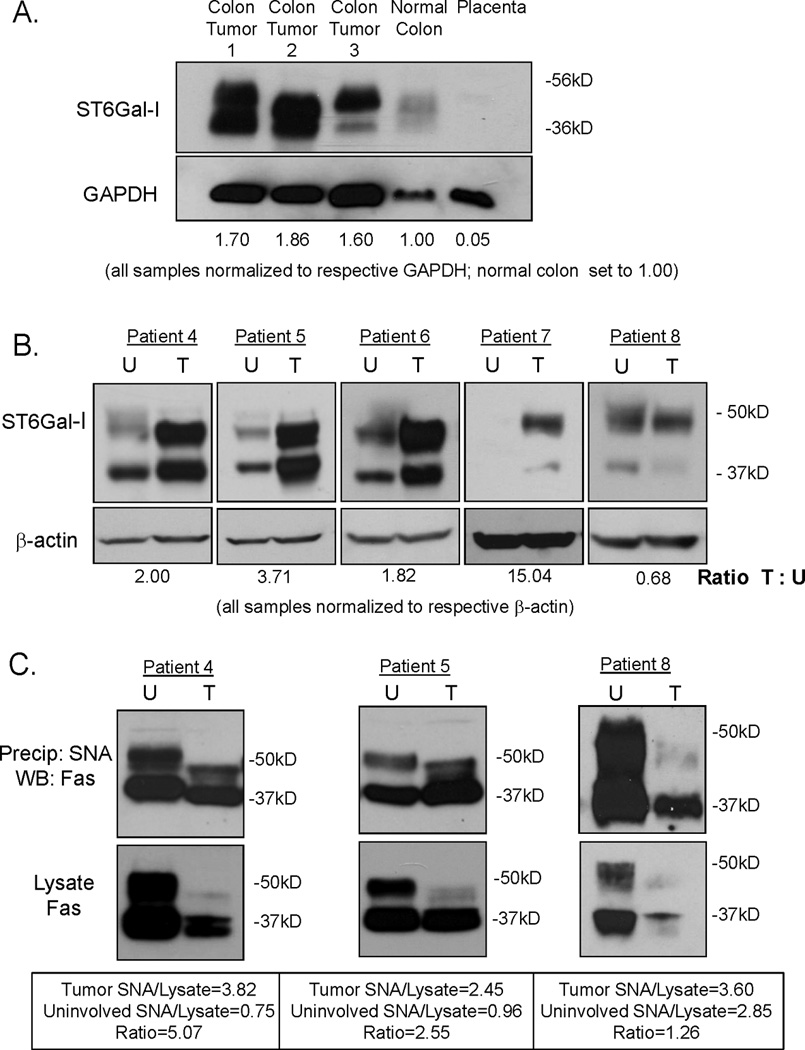
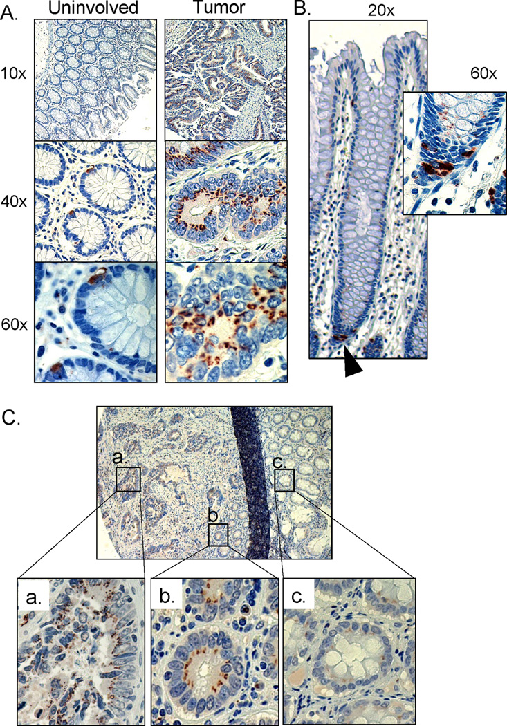
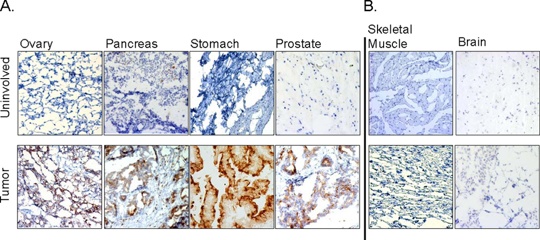
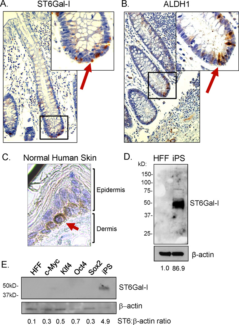
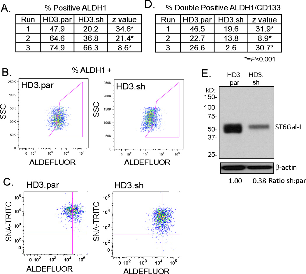
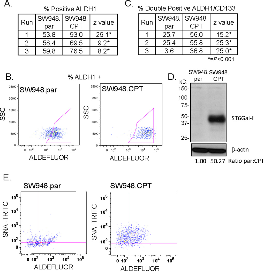
The ST6Gal-I sialyltransferase adds an α2-6-linked sialic acid to the N-glycans of certain receptors. ST6Gal-I mRNA has been reported to be upregulated in human cancer, but a prior lack of antibodies has limited immunochemical analysis of the ST6Gal-I protein. Here, we show upregulated ST6Gal-I protein in several epithelial cancers, including many colon carcinomas. In normal colon, ST6Gal-I localized selectively to the base of crypts, where stem/progenitor cells are found, and the tissue staining patterns were similar to the established stem cell marker ALDH1. Similarly, ST6Gal-I expression was restricted to basal epidermal layers in skin, another stem/progenitor cell compartment. ST6Gal-I was highly expressed in induced pluripotent stem (iPS) cells, with no detectable expression in the fibroblasts from which iPS cells were derived. On the basis of these observations, we investigated further an association of ST6Gal-I with cancer stem cells (CSC). Selection of irinotecan resistance in colon carcinoma cells led to a greater proportion of CSCs compared with parental cells, as measured by the CSC markers CD133 and ALDH1 activity (Aldefluor). These chemoresistant cells exhibited a corresponding upregulation of ST6Gal-I expression. Conversely, short hairpin RNA (shRNA)-mediated attenuation of ST6Gal-I in colon carcinoma cells with elevated endogenous expression decreased the number of CD133/ALDH1-positive cells present in the cell population. Collectively, our results suggest that ST6Gal-I promotes tumorigenesis and may serve as a regulator of the stem cell phenotype in both normal and cancer cell populations.


Conflict of interest statement

Authors have no conflict of interest to disclose.

Figures

Figure 1
Figure 1. ST6Gal-I is upregulated in human colon tumors and tumor-associated Fas receptors have elevated α2–6 sialylation
(A) A commercially purchased membrane for immunoblotting was probed for ST6Gal-I protein expression. Mature ST6Gal-I (upper band) is highly expressed in three separate colon tumors as compared to normal colon and placenta. The lower band is consistent with a cleaved, secreted form of ST6Gal-I (19). Densitometry was performed on the upper band. All samples were normalized to GAPDH and then compared to normal colon expression. (B) Tissue homogenates were prepared from colon tumors and pair-matched uninvolved colon specimens and immunoblotted for ST6Gal-I. ST6Gal-I was upregulated in 4/5 of colon tumors as compared to pair-matched uninvolved colon tissues. Densitometry was performed on the upper band. All samples were normalized to β-actin and then tumor samples were compared to pair-matched uninvolved colon. (C) α2–6 sialylated proteins were isolated using SNA1-agarose and immunoblotted for Fas. Total Fas levels were assessed by immunoblotting initial tissue homogenates (not subjected to SNA) for Fas. In all three patient samples, mature Fas (upper band) was downregulated in the tumor tissue relative to the respective uninvolved tissue. In patients with upregulated ST6Gal-I (patients 4 and 5), the proportion of sialylated Fas to total Fas was 5.07 and 2.55 respectively (densitometry was performed on the upper band). In the patient without upregulation of ST6Gal-I (patient 8), the proportion of sialylated Fas to total Fas was much lower at 1.26. U=uninvolved, T=tumor
Figure 2
Figure 2. ST6Gal-I upregulation and localization in human colon tumors
(A) Representative sample of pair-matched tissues stained for ST6Gal-I protein expression. Paraffin embedded specimens of uninvolved colon tissue and tumor tissues were immunohistologically stained for ST6Gal-I (brown), and counterstained with hematoxylin (blue). ST6Gal-I was highly upregulated in tumor tissue, whereas in uninvolved colon tissue, expression was restricted to a very few cells within each crypt structure. (B) Longitudinal view of a crypt from uninvolved tissue. ST6Gal-I staining was restricted to the base of the crypt (black arrow). Inset shows enlarged view with ST6Gal-I stain in cells at the base of the crypt. (C) ST6Gal-I staining in a patient sample showed gradient expression based on proximity to tumor. (a) upregulated expression of ST6Gal-I in malignant tissue, (b) aberrant expression in morphologically normal crypt structures directly adjacent to tumor and (c) low expression in crypts distal to the tumor.
Figure 3
Figure 3. ST6Gal-I is upregulated in several types of epithelial cancers, but not non-epithelial cancers
(A) Frozen epithelial tumors and pair-matched uninvolved tissues from ovary, pancreas, stomach, and prostate were stained for ST6Gal-I protein expression and counterstained with hematoxylin. ST6Gal-I upregulation was apparent in the tumor samples. (B) Frozen pair-matched tissues from skeletal muscle and brain exhibited low or undetectable levels of ST6Gal-I.
Figure 4
Figure 4. ST6Gal-I expression in stem/progenitor cell populations
(A) Paraffin-embedded normal colon tissue (from a cancer-free patient) was stained for ST6Gal-I. ST6Gal-I expression was confined to the base of normal colon crypts, with no expression observed in the apical, differentiated epithelium. (B) Expression of the ALDH1 stem cell marker was localized to the base of the crypts in normal human colon, similar to ST6Gal-I. (C) Staining for ST6Gal-I in paraffin-embedded normal human skin tissue. ST6Gal-I expression was confined to the basal proliferative compartment of the epidermis in normal skin. (D) Immunoblot for ST6Gal-I expression in cell lysates obtained from human induced Pluripotent Stem cells (iPS) and the human foreskin fibroblast population (HFF) from which iPS cells were derived. There was no detectable ST6Gal-I expression in HFFs, whereas there was a dramatic upregulation of ST6Gal-I in the iPS cells. (E) Immunoblot for ST6Gal-I expression in HFFs, iPS cells, or HFFs transduced with only one of the individual Yamanaka factors: c-Myc; Klf4; Oct4; or Sox2. ST6Gal-I upregulation was observed in iPS cells (generated by simultaneous transduction of all four factors), but not in HFFs transduced with the single factors alone. Densitometry was completed by normalizing signal to the respective β-actin band and then comparing HFFs to iPS.
Figure 5
Figure 5. ST6Gal-I expression correlated with cancer stem cell enrichment
(A) Colon carcinoma cells, HD3.par and HD3.sh, were assayed for ALDH1 activity (Aldefluor) by flow cytometry. Enrichment of ALDH1 staining was significantly higher in HD3.par as compared to HD3.sh in three independent runs. (B) Representative dot plot (run #1, 5A) showing ALDH1 staining. (C) Aldefluor and SNA-TRITC double-labeling shows knockdown decreases α2–6 surface sialylation along with stem cell enrichment. (D) Double labeling for stem cell enrichment of HD3.par and HD3.sh cells with ALDH1 and CD133 by flow cytometry revealed that knockdown of ST6Gal-I lead to significantly decreased enrichment in three independent runs. (E) Immunoblot of HD3.par and HD3.sh cells showed that shRNA transduction reduced ST6Gal-I expression. Densitometry completed by normalizing to respective β-actin and then comparing HD3.sh to HD3.par. *=P <0.001.
Figure 6
Figure 6
(A) ALDH1 activity was assayed by flow cytometry in colon carcinoma cell line SW948. SW948.CPT chemoresistant line had significant enrichment for ALDH1 staining in three independent runs as compared to SW948.par. (B) Representative dot plot of ALDH1 staining 28 (run #1, 6A). (C) Double-labeling of SW948.par and SW948.CPT with ALDH1 and CD133 showed significant increase in stem cell markers in the chemoresistant line (SW948.CPT) in three independent runs. (D) Immunoblot of SW948.par and SW948.CPT shows ST6Gal-I expression was upregulated in the SW948.CPT line. Densitometry completed by normalizing to respective β-actin and then comparing SW948.CPT to SW948.par. (E) Double-labeling with Aldefluor and SNA-TRITC demonstrates chemoresistant line has increased stem cell enrichment as well as increased surface α2–6 sialylation. *=P <0.001.

References

    1. Schultz MJ, Swindall AF, Bellis SL. Regulation of the metastatic cell phenotype by sialylated glycans. Cancer Metastasis Rev. 2012;31:501–518. - PMC - PubMed
    1. Zhuo Y, Bellis SL. Emerging role of alpha2,6-sialic acid as a negative regulator of galectin binding and function. J Biol Chem. 2010;286:5935–5941. - PMC - PubMed
    1. Dall'Olio F, Chiricolo M. Sialyltransferases in cancer. Glycoconj J. 2001;18:841–850. - PubMed
    1. Zhu Y, Srivatana U, Ullah A, Gagneja H, Berenson CS, Lance P. Suppression of a sialyltransferase by antisense DNA reduces invasiveness of human colon cancer cells in vitro. Biochim Biophys Acta. 2001;1536:148–160. - PubMed
    1. Lin S, Kemmner W, Grigull S, Schlag PM. Cell surface alpha 2,6 sialylation affects adhesion of breast carcinoma cells. Exp Cell Res. 2002;276:101–110. - PubMed

Publication types

MeSH terms