. 2013 May;34(5):1158-64.
doi: 10.1093/carcin/bgt021. Epub 2013 Jan 28.

Basal stem cells contribute to squamous cell carcinomas in the oral cavity

Affiliations

Basal stem cells contribute to squamous cell carcinomas in the oral cavity

Xiao-Han Tang et al. Carcinogenesis. 2013 May.

Abstract

The cells of origin of oral cavity squamous cell carcinoma (OCSCC) are unknown. We used a cell lineage tracing approach (adult K14-CreER(TAM); ROSA26 mice transiently treated with tamoxifen) to identify and track normal epithelial stem cells (SCs) in mouse tongues by X-gal staining and to determine if these cells become neoplastically transformed by treatment with a carcinogen, 4-nitroquinoline 1-oxide (4-NQO). Here, we show that in normal tongue epithelia, X-gal(+) cells formed thin columns throughout the entire epithelium 12 weeks after tamoxifen treatment, indicating that the basal layer contains long-lived SCs that produce progeny by asymmetric division to maintain homeostasis. Carcinogen treatment results in a ~10-fold reduction in the total number of X-gal(+) clonal cell populations and horizontal expansion of X-gal(+) clonal cell columns, a pattern consistent with symmetric division of some SCs. Finally, X-gal(+) SCs are present in papillomas and invasive OCSCCs, and these long-lived X-gal(+) SCs are the cells of origin of these tumors. Moreover, the resulting 4-NQO-induced tumors are multiclonal. These findings provide insights into the identity of the initiating cells of oral cancer.


Figures

Fig. 1.
Fig. 1.
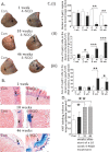
Epithelial basal SCs are present in normal mouse tongues. (A) Whole-mount X-gal staining. K14-CreERTAM; ROSA26 mouse tongues at various time points after the administration of tamoxifen (×8, the tip portions of mouse tongues are shown here). (B) H&E-stained sections from X-gal-stained tongues at various time points after the administration of tamoxifen. D: dorsal; V: ventral (×200; scale bars, 50 μm). X-gal staining is indicated by arrows (black or white).
Fig. 2.
Fig. 2.
The carcinogen, 4-NQO, reduces the numbers of X-gal(+) cell populations and increases the size of some X-gal(+) cell populations in tongues. X-gal staining is indicated by an arrow. In (A) and (B), Con, control; 4-NQO, 4-NQO treated. (A) Whole-mount X-gal staining on K14-CreERTAM; ROSA26 mouse tongues at various time points after the start of 4-NQO administration (×8, the tip portions of mouse tongues are shown here though we measured the dorsal surfaces of the entire tongues). For 1 and 10 weeks, n = 3 per treatment; for 46 weeks, n = 5 for control and n = 22 for 4-NQO. (B) H&E-stained sections from X-gal-stained tongues at various time points after the start of 4-NQO administration (×200; scale bars, 50 μm). (C) quantification of the numbers and areas of X-gal(+) cell populations. (i) The numbers of X-gal(+) cells on the dorsal side of tongues, (ii) the sizes of the biggest X-gal(+) cell populations on the dorsal side of mouse tongues and (iii) the sizes of the biggest X-gal(+) cell groups in H&E-stained sections (three mice at each time point and six to eight sections from each mouse). In each panel, C: control; N: 4-NQO treated. (D) Quantification of Ki67 labeling indices in tongue epithelia. Differences with a P value of <0.05 (marked with an asterisk), P < 0.01 (marked with two asterisks) and P < 0.001 (marked with three asterisks) after one-way analysis of variance and subsequently the Bonferroni test for multiple comparisons (two-tailed test) were considered to be statistically significant.
Fig. 3.
Fig. 3.
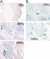
X-gal(+) cells are present in the papilloma in 4-NQO-treated tongues. Whole-mount X-gal staining was performed on the tongues prior to paraffin embedding, sectioning and H&E staining. (A) (i) Macroscopic picture of papilloma (indicated by arrows) in 4-NQO-treated mouse tongue (×8). (ii) H&E-stained tongue section showing the expansion of X-gal(+) cell populations (×40; scale bar, 200 μm). (iii) H&E-stained papillomas (arrows) shown in panel (i) (×40; scale bar, 200 μm). (iv and v) Higher magnifications of the papilloma shown in panel (iii) (×200; scale bar, 50 μm; ×600; scale bar, 20 μm, respectively); (B) Ki67 staining (indicated by an arrow) in the papilloma shown in panel (Aiv). Scale bars are 50 μm for ×200 and 20 μm for ×600.
Fig. 4.
Fig. 4.
X-gal(+) cells are present in the invasive SCCs in 4-NQO-treated tongues. Whole-mount X-gal staining was performed on the tongues prior to paraffin embedding. (A) H&E-stained invasive SCCs (×40; scale bar, 200 μm; ×200; scale bar, 50 μm; ×600; scale bar, 20 μm); (B) Ki67 staining (marked by an arrow) in the invasive SCC shown in panel (A). Scale bars are 50 μm for ×200 and 20 μm for ×600.
Fig. 5.
Fig. 5.
Model of homeostasis of tongue epithelia. Under normal conditions, tongue epithelial basal cells undergo ‘asymmetric divisions’ (red arrow) to both self-renew and produce a progeny cell destined for differentiation in the suprabasal layers, and some cells in the superficial layers are shed (asterisks); in 4-NQO-treated tongues, some basal SCs are killed by the carcinogen, and the surviving, neighboring basal SCs undergo ‘symmetric’ divisions (blue arrow) to compensate for the loss of SCs. In addition, they still undergo asymmetric divisions (red arrow) to self-renew and produce differentiated progeny in suprabasal layers. Blue cells, X-gal(+) cells; non-blue cells, X-gal(−) cells.

References

    1. Siegel R., et al. (2011). Cancer statistics, 2011: the impact of eliminating socioeconomic and racial disparities on premature cancer deaths. CA Cancer J. Clin., 61, 212–236 - PubMed
    1. Binnie W.H., et al. (1983). Etiology of oral squamous cell carcinoma. J. Oral Pathol., 12, 11–29 - PubMed
    1. Lippman S.M., et al. (2005). Oral cancer prevention and the evolution of molecular-targeted drug development. J. Clin. Oncol., 23, 346–356 - PubMed
    1. Kupferman M.E., et al. (2007). Molecular analysis of anoikis resistance in oral cavity squamous cell carcinoma. Oral Oncol., 43, 440–454 - PubMed
    1. Tsai L.L., et al. (2011). Markedly increased Oct4 and Nanog expression correlates with cisplatin resistance in oral squamous cell carcinoma. J. Oral Pathol. Med., 40, 621–628 - PubMed

Publication types

MeSH terms