Review
. 2013 May;29(5):849-54.
doi: 10.1007/s00381-013-2037-4. Epub 2013 Jan 29.

Embryonal tumor with abundant neuropil and true rosettes: an autopsy case-based update and review of the literature

Affiliations
Review

Embryonal tumor with abundant neuropil and true rosettes: an autopsy case-based update and review of the literature

Dariusz Adamek et al. Childs Nerv Syst. 2013 May.

Abstract

Introduction: Embryonal tumor with abundant neuropil and true rosettes (ETANTR) is a rare subtype of primitive neuroectodermal tumors first reported in 2000. It is rare among the group of embryonal central nervous system tumors with approximately 50 reported cases. ETANTR has been suggested to be a separate entity among this group of tumors.

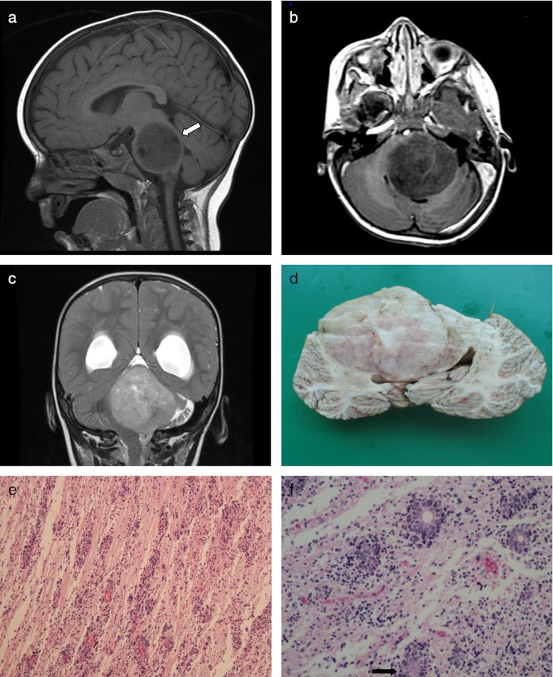
Case report: Herein, we present only the second autopsy case of ETANTR, which occurred in a 17-month-old boy, and was located in the brainstem. The tumor was inoperable, and despite chemotherapy, the child died 3 months after initial hospitalization. A brain only autopsy was performed.

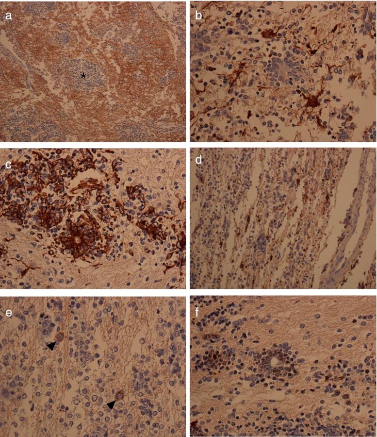
Discussion: Neuropathological and neuroimaging examinations suggest ETANTR grew by expansion rather than invasion distorting anatomical structures of the posterior fossa. We suggest that the characteristic histopathological picture of the tumor is the result of multifocal and dispersed germinative activity surrounded by mature neuropil-like areas.

Conclusion: ETANTR is a pediatric tumor occurring in children under 4 with a significantly poor prognosis with more cases and research required to characterize this rare embryonal tumor.


Figures

Fig. 1
Fig. 1
a MRI, 3DFSPGRT1 sequence, sagittal plane. The tumor is localized in the brainstem and is extremely expanding and deforming its structure. Noteworthy is severe compression of the fourth ventricle, but evidently between the tumor and lumen of the fourth ventricle, there remains a thin, uninterrupted strip of brain tissue (arrow). b MRI, DGRET1 with contrast, axial plane. Contrast enhancement within the tumor is conspicuously insignificant. c MRI, frFSET2, coronal plane. The tumor appears as a very well delineated expansive process, with heterogeneous signal, containing some fluid-filled areas at the level of fourth ventricle and medial part of left cerebellar hemisphere, which has atrophied. The tumor molds and compresses the brainstem and obliterates the fourth ventricle. Supratentorially, there are features of hydrocephalus with transudate of CSF into surrounding brain tissue. d Macroscopic cross section of the tumor at the level of the cerebellum. Noteworthy is the very distinct border between the tumor and apparently untouched but evidently severely displaced and compressed cerebellar tissue. e Characteristic mixture of neuropil-like paucicellular zones with quite uniformly scattered cellular “densities” consisting of small undifferentiated cells. Both within densities and also directly within neuropil zones, one could discern rosettes which are quite characteristic for this tumor, H&E, objective magnification ×10. f A close-up view of rosettes. Some with empty core but delicately contoured with pinkish line, and some with homogenous featureless core (arrow), H&E, objective magnification ×40
Fig. 2
Fig. 2
a The more cellular zones (asterisk), in contrary to neuropil background, are rather negative for synaptophysin, objective magnification ×20. b All rosettes were negative for synaptophysin. Apart from the rosettes, strongly synaptophysin-positive cells with prominent processes resembling astrocytes rather than neurons were noted, objective magnification x40. c Both rosettes and almost all cells within cellular “densities” are strongly immunopositive for vimentin, objective magnification ×40. d Rosettes are negative for S100. However, between the rosettes there are S-100 positive cells with marked processes, objective magnification ×20. e Some mature looking synaptophysin-positive neurons (arrowheads) are scattered within neuropil, objective magnification ×40. f Proliferating activity as evidenced by immunopositivity to Ki67 was marked within rosettes and also around vessels (not shown in this picture), objective magnification ×40

References

    1. Al-Hussain TO, Dababo MA. Posterior fossa tumor in a 2-year-old girl. Brain Pathol. 2009;19:343–346. doi: 10.1111/j.1750-3639.2009.00279.x. - DOI - PMC - PubMed
    1. Biegel JA, Allen CS, Kawasaki K, Shimizu N, Budarf ML, Bell CJ. Narrowing the critical region for a rhabdoid tumor locus in 22q11. Genes Chromosomes Cancer. 1996;16:94–105. doi: 10.1002/(SICI)1098-2264(199606)16:2<94::AID-GCC3>3.0.CO;2-Y. - DOI - PubMed
    1. Buccoliero AM, Castiglione F, Rossi Degl'Innocenti D, Franchi A, Paglierani M, Sanzo M, Cetica V, Giunti L, Sardi I, Genitori L, Taddei GL. Embryonal tumor with abundant neuropil and true rosettes: morphological, immunohistochemical, ultrastructural and molecular study of a case showing features of medulloepithelioma and areas of mesenchymal and epithelial differentiation (case report) Neuropathology. 2010;30:84–91. doi: 10.1111/j.1440-1789.2009.01040.x. - DOI - PubMed
    1. Dunham C, Sugo E, Tobias V, Wills E, Perry A. Embryonal tumor with abundant neuropil and true rosettes (ETANTR): report of a case with prominent neurocystic differentiation. J Neurooncol. 2007;84:91–98. doi: 10.1007/s11060-007-9346-y. - DOI - PubMed
    1. Eberhart CG, Brat DJ, Cohen KJ, Burger PC. Pediatric neuroblastic brain tumors containing abundant neuropil and true rosettes. Pediatr Dev Pathol. 2000;3:346–352. doi: 10.1007/s100249910049. - DOI - PubMed

LinkOut - more resources