. 2013 Feb 11;210(2):241-56.
doi: 10.1084/jem.20121977. Epub 2013 Jan 28.

Identification of autoantigens recognized by the 2F5 and 4E10 broadly neutralizing HIV-1 antibodies

Affiliations

Identification of autoantigens recognized by the 2F5 and 4E10 broadly neutralizing HIV-1 antibodies

Guang Yang et al. J Exp Med. .

Abstract

Many human monoclonal antibodies that neutralize multiple clades of HIV-1 are polyreactive and bind avidly to mammalian autoantigens. Indeed, the generation of neutralizing antibodies to the 2F5 and 4E10 epitopes of HIV-1 gp41 in man may be proscribed by immune tolerance because mice expressing the V(H) and V(L) regions of 2F5 have a block in B cell development that is characteristic of central tolerance. This developmental blockade implies the presence of tolerizing autoantigens that are mimicked by the membrane-proximal external region of HIV-1 gp41. We identify human kynureninase (KYNU) and splicing factor 3b subunit 3 (SF3B3) as the primary conserved, vertebrate self-antigens recognized by the 2F5 and 4E10 antibodies, respectively. 2F5 binds the H4 domain of KYNU which contains the complete 2F5 linear epitope (ELDKWA). 4E10 recognizes an epitope of SF3B3 that is strongly dependent on hydrophobic interactions. Opossums carry a rare KYNU H4 domain that abolishes 2F5 binding, but they retain the SF3B3 4E10 epitope. Immunization of opossums with HIV-1 gp140 induced extraordinary titers of serum antibody to the 2F5 ELDKWA epitope but little or nothing to the 4E10 determinant. Identification of structural motifs shared by vertebrates and HIV-1 provides direct evidence that immunological tolerance can impair humoral responses to HIV-1.


Figures

Figure 1.
Figure 1.
Identification of 2F5 and 4E10 cellular antigens by immunoprecipitation. (A) HEp-2 cell lysate was immunoprecipitated with 2F5, 4E10, or human myeloma IgG (151K) and resolved under nonreducing conditions as described in Materials and methods. Boxes indicate bands that were differentially associated with 2F5, 4E10, or 151K eluates and were excised in pairs (BnAb versus 151K) for mass spectrometric analysis. HEp-2 lane contains whole-cell lysate of HEp-2; 2F5 IP, 151K IP, and 4E10 IP lanes include specifically bound proteins eluted from 2F5, 151K, and 4E10, respectively. Protein molecular masses (kD) are indicated on both sides of gel electrophoresis. The experiment was repeated on four independent occasions. (B) Excised protein bands were analyzed by tandem mass spectrometry; candidates were defined as proteins uniquely recovered in the BnAb, but not 151K control bands of the same molecular masses. To identify immunoprecipitates accurately, we required each protein candidate to be represented by three or more peptides with >95% peptide identification probability. Listed proteins were recovered in two or more out of three independent mass spectrometry tryptic digest analyses. Protein molecular mass indicates size of the band from which proteins were identified.
Figure 2.
Figure 2.
Identification of 2F5 and 4E10 self-antigens by protein array. Representative ProtoArray summary for protein arrays blotted with 2F5 (A), 4E10 (B), or 151K control. Axis values are relative fluorescence signal intensity in 151 array (y-axis) or BnAb array (x-axis). All proteins appear as duplicates on each array and each dot represents one replicate. Diagonal indicates equal binding by BnAb and 151K. Circles indicate repeatedly recovered positive hits by each Ab. BHMT2 (betaine-homocysteine methyltransferase 2) and COX7A2L (cytochrome c oxidase subunit VIIa polypeptide 2 like) were identified as the specific ligand for the human myeloma 151K in four or more independent experiments. As a control, we compared the binding pattern of a humanized therapeutic mAb against TNF, Infliximab, to that of the myeloma protein 151K (not depicted). Infliximab strongly associated with TNF, significantly better than the 151K, confirming that our system is capable of identifying the specific ligand of an Ab. IL1F6, IL-1 family member 6; Rxr-β, retinoid X receptor β. 2F5 and 151K arrays were repeated in three or more independent occasions; 4E10 protein array was performed twice.
Figure 3.
Figure 3.
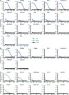
Screen of candidates for 2F5 and 4E10 self-ligands. Serial dilutions of 2F5 (A and B, blue), 4E10 (C and D, green), and 151K (black) in duplicates were incubated on ELISA plates preimmobilized with the recombinant candidate proteins identified in Figs. 1 and 2. BSA was used as negative control. The first round of screen was performed under permissive binding conditions as described in Materials and methods (A and C). Seven strongly recognized ligands for 2F5 and all 4E10 candidate ligands were then subject to a second round of screen under stringent binding conditions as described in Materials and methods (B and D). Axis values are OD (y-axis) or antibody concentrations (ng/ml; x-axis). All experiments were repeated in two or more independent experiments.
Figure 4.
Figure 4.
KYNU and SF3B3 are the main self-ligands of 2F5 and 4E10. Inhibition of 2F5 binding to immobilized recombinant KYNU (A) or CMTM3 (B) assessed in ELISA as described in Materials and methods. The y-axis indicates OD percentage of maximal binding, which is determined as the mean reading without inhibitors (100% binding is marked by gray dashes for comparison). The x-axis designates the molar ratio of inhibitor to plate immobilized ligand. (C) 2F5 and 151K immunoprecipitation eluates were stained with KYNU Ab in Western blot. Recombinant KYNU (rKYNU) was used as a positive control; molecular masses (kD) of protein markers are shown on the left of gel. (D and E) HEp-2 slides were indirectly labeled with anti-KYNU (FITC, green) and 2F5 (PE, red; D) or 4E10 (FITC, green) and anti-SF3B3 (PE, red; E). Negative controls were included to ensure specific staining and minimal cross-reactivity of all primary and secondary Abs. Bars, 15 µm. All data are representative of three or more independent experiments.
Figure 5.
Figure 5.
2F5 recognizes KYNU through the H4 DKW shared with HIV-1 gp41. (A) Structure of monomeric KYNU as it appears in the dimer complex. Green highlights 2F5 epitope within the H4 domain of KYNU; red indicates D amino acid in the core of 2F5 epitope (Lima et al., 2007). (B) Recombinant wild-type and mutant KYNU were resolved in nonreducing SDS-PAGE and stained with coomassie blue with molecular mass standards (KD) on the left. (C and E) gp41, recombinant KYNU, and mutant KYNU binding to anti-KYNU (C) or 2F5 (E) in Luminex assays. Uncoupled beads (blank) were used as negative control. MFI, mean fluorescence intensity. (D) Enzymatic activities of wild-type and mutant KYNU were measured as described in Materials and methods. RFU, relative fluorescence unit. Enzymatic measurements were repeated in three or more independent experiments. All other data were collected twice in duplicates.
Figure 6.
Figure 6.
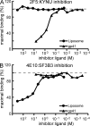
4E10 binds SF3B3 via hydrophobic interactions. Competitive inhibition of 2F5 binding to KYNU (A), and 4E10 binding to SF3B3 (B) by gp41 (triangle) or liposome containing 75% cardiolipin and 25% phosphatidylcholine (circle). Recombinant KYNU and SF3B3 were immobilized, and threefold serial dilutions of the indicated inhibitor were added, followed by 200 ng/ml 2F5 (A) or 4E10 (B). BnAb binding was detected as described in Materials and methods. The y-axis indicates OD percentage of maximal binding, which is determined as the mean reading without inhibitors (100% binding is marked by gray dashes for comparison). The x-axis designates the molar ratio of inhibitor to plate immobilized ligand. All experiments were performed in duplicates.
Figure 7.
Figure 7.
KYNU and SF3B3 are recognized by germline precursors of 2F5 and 4E10. (A and B) Recombinant KYNU (A) or mutant KYNU (B) were immobilized on ELISA plates and detected by duplicate threefold serial dilutions of 2F5, 2F5 RUA variant 1, 2F5 RUA variant 2, and anti-KYNU. (C) Recombinant SF3B3 was immobilized, duplicate serial dilutions of 4E10, 4E10 GL1, and anti-SF3B3 were added, and binding was detected as described in Materials and methods. All experiments include negative controls with BSA plates and isotype control Ab (not depicted). All data are representative of at least three independent experiments.
Figure 8.
Figure 8.
KYNU, CMTM3, and SF3B3 are phylogenetically conserved. (A) Amino acid sequence flanking the 2F5 epitope within the MPER of HIV-1 gp41. (B) The amino acid sequences of the H4 domain of KYNU in human, chimpanzee, mice, and opossums are listed. (C) CMTM3 sequence flanking DKW in human, chimpanzee, mice, and opossums. (D) Overall amino acid sequence identity of mammalian SF3B3 compared with human SF3B3. Sequences of all mammalian species for which the genome has been sequenced are also conserved in the 2F5 epitope in KYNU (not depicted).
Figure 9.
Figure 9.
Opossums mount robust 2F5-like Ab responses after immunization with HIV-1 envelope protein. (A) 12 laboratory opossums were immunized with JR-FL gp140 and boosted with liposomes containing MPER peptide and TLR ligands. MPER peptide contains both 2F5 and 4E10 determinants. Sera were assayed by ELISA for opossum IgG against gp41, biotinylated 2F5-epitope peptide, and biotinylated 4E10 epitope peptide. All ELISA plates contained positive controls of 2F5 or 4E10 and negative controls of human IgG (not depicted). End-point titers (n = 12; geometric means ± SD) were determined as described in Materials and methods. For comparison, BL/6 mice were primed and boosted i.p. with the same JR-FL gp140 and liposome immunogens given to opossums at 2-wk intervals for 12 wk. Mouse sera (pre and postimmunization) were assayed by ELISA for IgG Ab using the MPER656 peptide that includes both the 2F5 and 4E10 epitopes. End-point titers (n = 5; geometric means ± SD) were determined as for opossums. (B) Single alanine mutations were introduced into a collection of 2F5 epitope peptides spanning residues 661–669. The effects of these mutations were evaluated by SPR and compared with binding of opossum serum to wild-type 2F5 peptide. The five sera with highest SP62 responses in SPR are shown.

References

    1. Adams E., Basten A., Goodnow C.C. 1990. Intrinsic B-cell hyporesponsiveness accounts for self-tolerance in lysozyme/anti-lysozyme double-transgenic mice. Proc. Natl. Acad. Sci. USA. 87:5687–5691 10.1073/pnas.87.15.5687 - DOI - PMC - PubMed
    1. Alam S.M., McAdams M., Boren D., Rak M., Scearce R.M., Gao F., Camacho Z.T., Gewirth D., Kelsoe G., Chen P., Haynes B.F. 2007. The role of antibody polyspecificity and lipid reactivity in binding of broadly neutralizing anti-HIV-1 envelope human monoclonal antibodies 2F5 and 4E10 to glycoprotein 41 membrane proximal envelope epitopes. J. Immunol. 178:4424–4435 - PMC - PubMed
    1. Alam S.M., Scearce R.M., Parks R.J., Plonk K., Plonk S.G., Sutherland L.L., Gorny M.K., Zolla-Pazner S., Vanleeuwen S., Moody M.A., et al. 2008. Human immunodeficiency virus type 1 gp41 antibodies that mask membrane proximal region epitopes: antibody binding kinetics, induction, and potential for regulation in acute infection. J. Virol. 82:115–125 10.1128/JVI.00927-07 - DOI - PMC - PubMed
    1. Alam S.M., Morelli M., Dennison S.M., Liao H.X., Zhang R., Xia S.M., Rits-Volloch S., Sun L., Harrison S.C., Haynes B.F., Chen B. 2009. Role of HIV membrane in neutralization by two broadly neutralizing antibodies. Proc. Natl. Acad. Sci. USA. 106:20234–20239 10.1073/pnas.0908713106 - DOI - PMC - PubMed
    1. Alam S.M., Liao H.X., Dennison S.M., Jaeger F., Parks R., Anasti K., Foulger A., Donathan M., Lucas J., Verkoczy L., et al. 2011. Differential reactivity of germ line allelic variants of a broadly neutralizing HIV-1 antibody to a gp41 fusion intermediate conformation. J. Virol. 85:11725–11731 10.1128/JVI.05680-11 - DOI - PMC - PubMed

Publication types

MeSH terms