. 2013:980:157-73.
doi: 10.1007/978-1-62703-287-2_8.

Quantitative real-time PCR expression analysis of peripheral blood mononuclear cells in pancreatic cancer patients

Affiliations

Quantitative real-time PCR expression analysis of peripheral blood mononuclear cells in pancreatic cancer patients

Michael J Baine et al. Methods Mol Biol. 2013.

Abstract

The ability of peripheral blood mononuclear cells (PBMCs) to act as a surrogate window into the presence and physiologic effects of pancreatic cancer is becoming increasingly apparent. In this chapter, we describe the techniques for isolation, lysis, RNA extraction, cDNA synthesis, and Q-RT PCR analysis of PBMCs as well as reasonable alternatives and the advantages and disadvantages of each. We further discuss the noteworthy considerations necessary for successful isolation and conversion of the high-quality PBMC RNA required to acquire interpretable and reproducible results for PBMC genetic expression analysis.


Figures

Fig. 1
Fig. 1
Layering of blood as visualized following ficoll density gradient centrifugation. Upon centrifugation of diluted whole blood layered over ficoll, as described, separation of various components occurs based on density. In total, four layers should be seen, with the aggregated erythrocytes forming a dark bottom layer, followed by the ficoll, then a thin, white layer of PBMCs, and finally the top layer of diluted plasma. Often, the PBMC layer is difficult to visualize but can be assumed to be present at the interface of the plasma and ficoll layers. In this case, PBMCs should be isolated by placing the tip of the pipette approximately 1 mm above the plasma/ficoll interface. Care should be taken not to disrupt the erythrocyte layer upon removal of the PBMCs or further RBC lysis will be necessary.
Fig. 2
Fig. 2
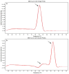
Melt curve analyses of Q-RT PCR results. Specificity of Q-RT PCR primers used with SYBR-Green-based chemistry can be easily accessed via melt curve analysis. As primer dimers and off-target primer associations will have different melting temperatures than the target amplified sequence, lack of primer specificity can be visualized as multiple peaks on melt curve. (a) A melt curve analysis showing a single peak, indicating primer specificity and valid interpretability of results. (b) Melt curve analysis showing two peaks, signifying off-target or primer-dimer associations of the utilized primers, preventing valid expression level quantification derived from the Q-RT PCR analysis. In the case of this, new primers should be designed for the Q-RT PCR analysis with greater care taken for sequence specificity using BLAST software.

References

    1. Baine MJ, Chakraborty S, Smith LM, et al. Transcriptional profiling of peripheral blood mononuclear cells in pancreatic cancer patients identifies novel genes with potential diagnostic utility. PLoS One. 2011;6:e17014. - PMC - PubMed
    1. Chakraborty S, Baine MJ, Sasson AR, Batra SK. Current status of molecular markers for early detection of sporadic pancreatic cancer. Biochim Biophys Acta. 2011;1815:44–64. - PMC - PubMed
    1. [This was accessed on December 21 2011];Surveillance Epidemiology, and End Results (SEER) Program. 2011 ( http://www.seer.cancer.gov)
    1. Bustin SA, Benes V, Garson JA, et al. The MIQE guidelines: minimum information for publication of quantitative real-time PCR experiments. Clin Chem. 2009;55:611–622. - PubMed

Publication types

MeSH terms