. 2013;8(1):e54663.
doi: 10.1371/journal.pone.0054663. Epub 2013 Jan 24.

The small GTPase Rif is dispensable for platelet filopodia generation in mice

Affiliations

The small GTPase Rif is dispensable for platelet filopodia generation in mice

Robert Goggs et al. PLoS One. 2013.

Abstract

Background: Formation of filopodia and other shape change events are vital for platelet hemostatic function. The mechanisms regulating filopodia formation by platelets are incompletely understood however. In particular the small GTPase responsible for initiating filopodia formation by platelets remains elusive. The canonical pathway involving Cdc42 is not essential for filopodia formation in mouse platelets. The small GTPase Rif (RhoF) provides an alternative route to filopodia generation in other cell types and is expressed in both human and mouse platelets.

Hypothesis/objective: We hypothesized that Rif might be responsible for generating filopodia by platelets and generated a novel knockout mouse model to investigate the functional role of Rif in platelets.

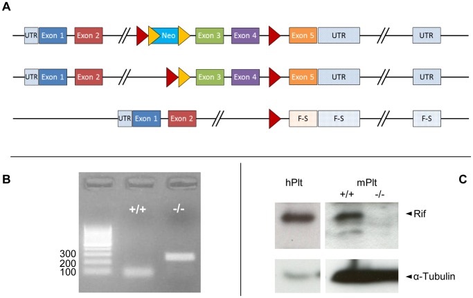
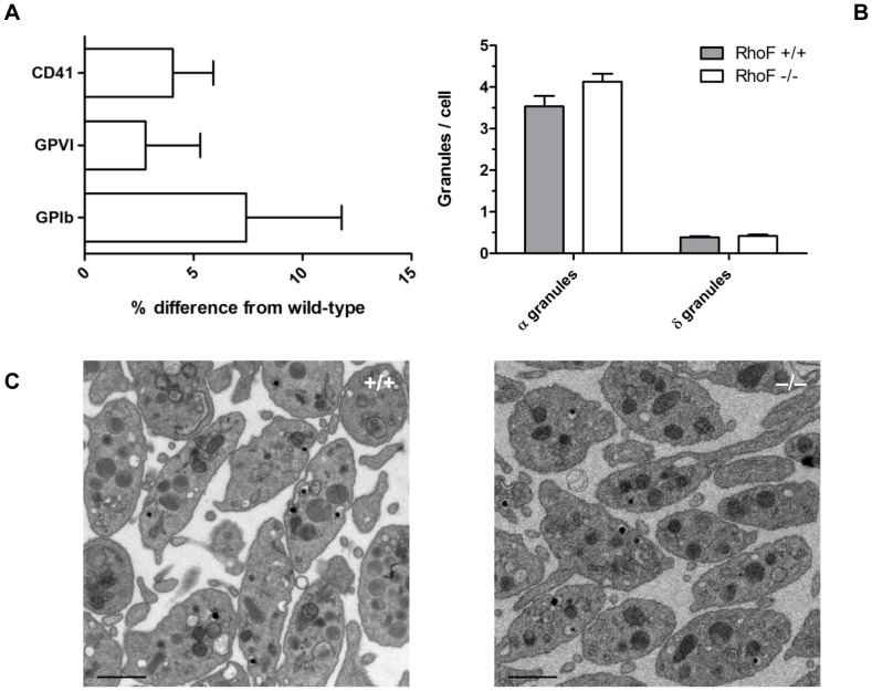
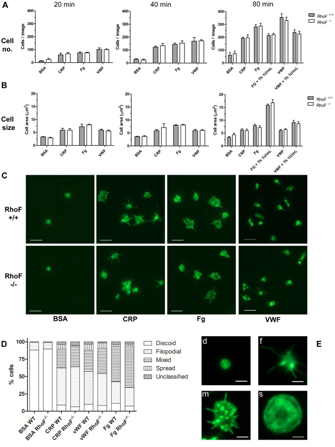
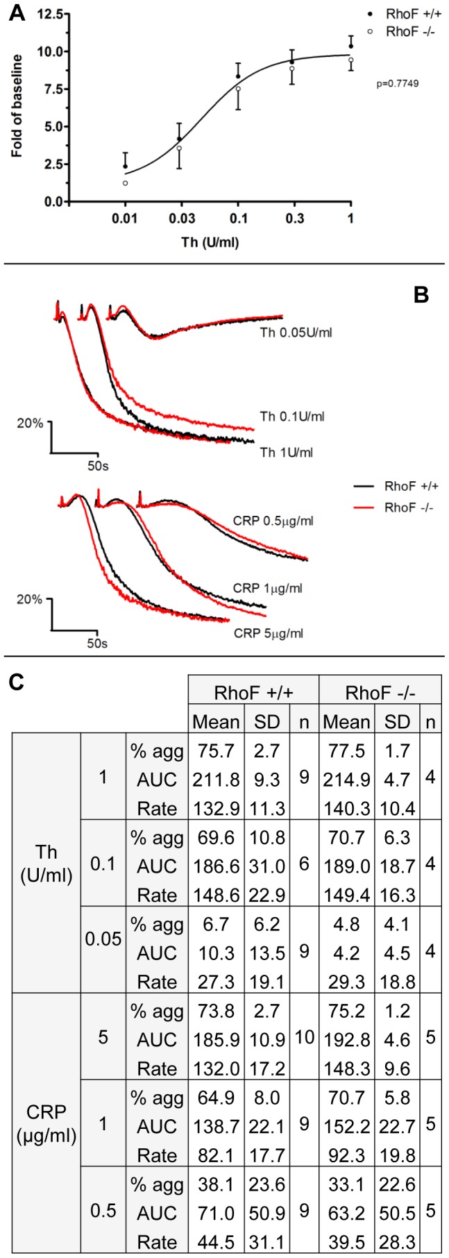
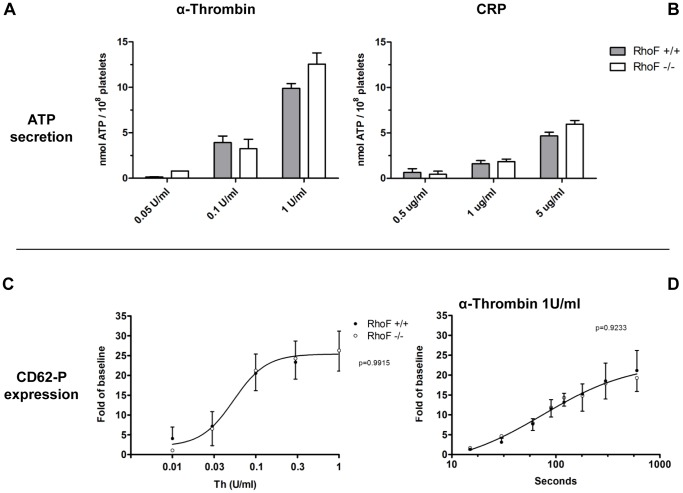
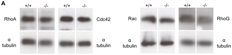
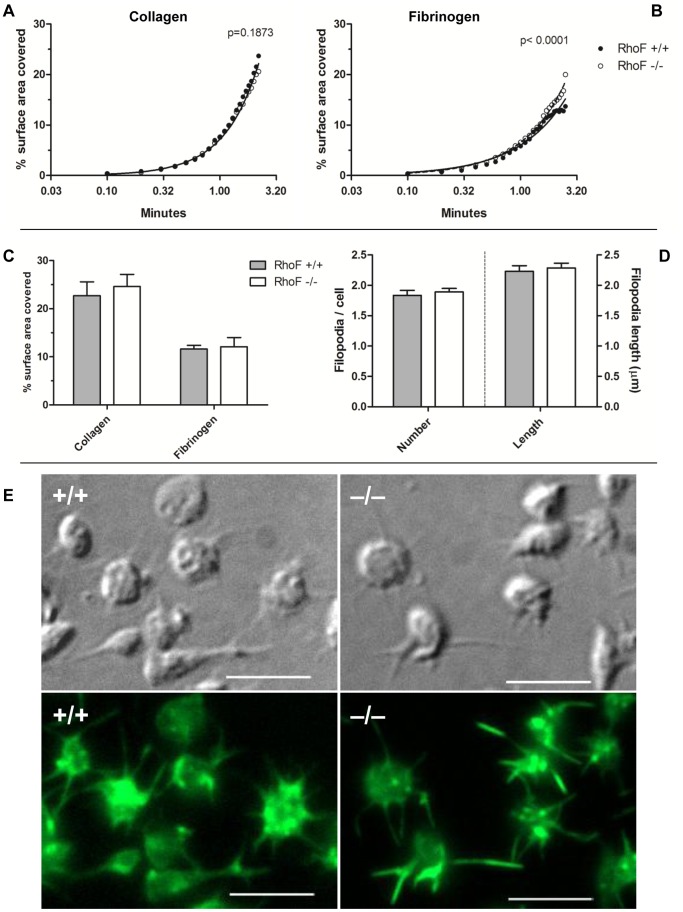
Methodology/principal findings: Constitutive RhoF(-/-) mice are viable and have normal platelet, leukocyte and erythrocyte counts and indices. RhoF(-/-) platelets form filopodia and spread normally on various agonist surfaces in static conditions and under arterial shear. In addition, RhoF(-/-) platelets have normal actin dynamics, are able to activate and aggregate normally and secrete from alpha and dense granules in response to collagen related peptide and thrombin stimulation.

Conclusions: The small GTPase Rif does not appear to be critical for platelet function in mice. Functional overlap between Rif and other small GTPases may be responsible for the non-essential role of Rif in platelets.


Conflict of interest statement

Competing Interests: Harry Mellor is a PLOS ONE Editorial Board member. The authors’ confirm that this does not alter their adherence to all the PLOS ONE policies on sharing data and materials.

Figures

Figure 1
Figure 1. Rif is expressed in human and mouse platelets and is ablated in platelets from a constitutive RhoF−/− mouse.
(A) Generation of the RhoF−/− mouse employed a targeting strategy which permitted generation of conditional or constitutive knockout (KO) mice. Exons 3 and 4 of the RhoF gene were flanked by LoxP sites and a Neo selection marker was inserted flanked by Frt sites such that deletion of exons 3 and 4 caused a frameshift mutation resulting in loss of function. Initially a conditional ready KO was generated after in vivo Flp-mediated removal of the selection marker. A constitutive KO was then generated after Cre-mediated deletion of exons 3 and 4. Red triangles represent LoxP sites, yellow triangles represent Frt sites. Abbreviations: untranslated region (UTR), frameshift (F-S). (B) The RhoF−/− mice genotyping strategy employed a three-primer duplex PCR reaction which generated a wild-type (+/+) product 100 bp long and a RhoF−/− product 250 bp long and enabled identification of both wild-type and RhoF−/− mice. (C) Analysis of Rif expression in human platelets (hPlt) and in wild-type (RhoF+/+) and Rif null (RhoF−/−) mouse platelets (mPlt) by Western blot demonstrating expression in human and wild-type mouse platelets and ablation of Rif protein expression in platelets from RhoF−/− mice.
Figure 2
Figure 2. Platelets from RhoF−/− mice have normal surface glycoprotein expression, normal granule numbers and normal ultrastructure.
(A) Expression of surface glycoproteins on RhoF−/− platelets was quantified by flow cytometry. Washed platelets were incubated with FITC labelled antibodies at saturating concentrations for 10 minutes at room temperature, then fixed and analysed by flow cytometry. Data are presented as mean ± SEM of the percentage difference from wild-type controls (n = 16). (B) Quantification of alpha and dense granule numbers per cell per thin section examined. Data are presented as mean ± SEM of 3 mice per group. (C) Representative transmission electron microscopy images of resting wild-type and RhoF−/− platelets. Scale bars represent 1 µm.
Figure 3
Figure 3. Platelets from RhoF−/− mice adhere to and spread on agonist surfaces under static conditions normally.
(A) Aliquots of washed platelets were allowed to adhere to and spread on fibrinogen (Fg), collagen related peptide (CRP) and von Willebrand Factor (VWF) coated surfaces. For VWF coated surfaces, platelets were allowed to adhere in the presence of 5 µg/ml botrocetin. Fatty-acid free BSA coated surfaces were used as negative controls. Co-stimulation with α-thrombin (1 U/ml) was also undertaken as indicated. Numbers of cells adherent per x63 field of view were quantified at the three indicated time points. (B) The average area of at least 500 adherent cells was quantified per agonist surface and 100 cells quantified per control. Data are presented as mean ± SEM of at least 4 mice per group. (C) Representative images of adherent cells on agonist and control surfaces at 20 minutes. Scale bars represent 5 µm. (D) Platelet morphology at 20 minutes was visually assessed and 100 cells per experiment assigned to one of 5 categories (discoid, filopodial, mixed, spread and unclassified). Data are presented as stacked bars representing the mean percentages of cells in each category from at least 4 mice per group. (E) Example images of discoid (d), filopodial (f), mixed (m) and spread (s) platelets corresponding to the morphology classifications represented in Fig. 3D. Unclassified cells were those platelets with morphologies that could not be assigned to any of these four groups. Scale bars represent 2 µm.
Figure 4
Figure 4. Integrin activation and light transmission aggregometry of RhoF−/− platelets is normal in response to thrombin and CRP.
(A) Dose-response curves for activation of integrin αIIbβ3 in response to thrombin stimulation as indicated by binding of the activation state specific antibody JON/A. No significant differences were identified between the fitted curves of wild-type and RhoF−/− platelets, suggesting that inside-out signalling to the integrin downstream of PARs does not require Rif. Data are presented as mean ± SEM of at least 5 mice per group. (B) Light transmission aggregation of RhoF−/− platelets in response to CRP is normal over a range of concentrations indicating that Rif is not required for signalling downstream of GPVI in mice. Similarly the aggregation responses of RhoF−/− platelets following thrombin stimulation are not significantly different from those of wild-type controls. Aggregation traces are representative of at least 4 mice per group. (C) Summary aggregation data comparing the three derived aggregation parameters, maximum aggregation at 3 minutes (% agg); the area under the aggregation curve to 3 minutes (AUC) and the maximum rate of aggregation (Rate) for RhoF−/− platelets and wild-type controls.
Figure 5
Figure 5
Release of ATP is normal in RhoF−/− mice in response to thrombin (A) and CRP (B) showing that Rif is not required for dense granule secretion. Data are presented as mean ± SEM of at least 4 mice per group. Similarly, expression of P-selectin induced by thrombin stimulation is normal over a range of concentrations (C) and follows a similar time course to that in wild-type controls (D). Rif does not therefore appear to be required for the cytoskeletal rearrangements required for alpha granule secretion in mouse platelets. Data are presented as mean ± SEM of at least 3 mice per group.
Figure 6
Figure 6. Expression of other small GTPase proteins is normal in RhoF−/− mice.
(A) Analysis of expression of the small GTPases Rac, RhoA, Cdc42 and RhoG in platelets from RhoF−/− mice. α-tubulin was used as a loading control.
Figure 7
Figure 7. Analyses of in vitro flow assay video frames at 6 second intervals.
Data points represent mean values of at least 8 mice per group. Analysis of adherence of platelets to immobilized collagen (A) indicates that accumulation of RhoF−/− platelets on collagen follows a similar time course to wild-type controls, suggesting that Rif is not required for initial adhesion events to collagen. (B) Video analyses of in vitro flow over fibrinogen identified a difference in the pattern of RhoF−/− platelet accumulation compared with wild type controls. (C) After a 3-minute wash at 1000 s−1 with buffer to remove erythrocytes and non-stably adhered platelets, 30 random images were taken and the surface area covered by platelets analysed. RhoF−/− mice form stable thrombi on both collagen and fibrinogen surfaces to a comparable degree to wild-type controls. Data are presented as mean ± SEM of at least 8 mice per group. (D) RhoF−/− platelets form actin-containing filopodia on fibrinogen surfaces under arterial shear conditions comparably with wild-type controls. Following in vitro flow of whole blood over fibrinogen coated surfaces, the number of filopodia per platelet and the length of filopodia were analyzed on 100 cells. Values are expressed as mean filopodia per cell and mean filopodia length ± SEM of 3 mice per group. (E) Post-washing, adherent cells were fixed with 500 µl 4% PFA flowed over surfaces at 1000 s−1, coverslips removed from flow chambers, permeabilized, stained with FITC-phalloidin and imaged by fluorescence and DIC microscopy. Identical parts of the images in both DIC and fluorescence channels are presented such that cell surface structures and the actin cytoskeleton within them can be directly compared. Scale bars represent 5 µm.

References

    1. Hartwig JH (2007) The platelet cytoskeleton. In: Michelson AD, editor. Platelets. 2 ed: Academic Press. 75–98.
    1. Eliautou S, Mangin PH, Walter U, Gachet C, Lanza F (2009) Normal filopodia extension in VASP-deficient platelets upon activation by adhesive matrices or soluble agonists. Thromb Haemost 102: 792–794. - PubMed
    1. Leng L, Kashiwagi H, Ren XD, Shattil SJ (1998) RhoA and the function of platelet integrin alphaIIbbeta3. Blood 91: 4206–4215. - PubMed
    1. McCarty OJ, Larson MK, Auger JM, Kalia N, Atkinson BT, et al. (2005) Rac1 is essential for platelet lamellipodia formation and aggregate stability under flow. J Biol Chem 280: 39474–39484. - PMC - PubMed
    1. Chang JC, Chang HH, Lin CT, Lo SJ (2005) The integrin alpha6beta1 modulation of PI3K and Cdc42 activities induces dynamic filopodium formation in human platelets. J Biomed Sci 12: 881–898. - PubMed

Publication types