. 2013 Apr 15;591(8):2205-19.
doi: 10.1113/jphysiol.2012.250852. Epub 2013 Jan 28.

Genetic ablation of aquaporin-2 in the mouse connecting tubules results in defective renal water handling

Affiliations

Genetic ablation of aquaporin-2 in the mouse connecting tubules results in defective renal water handling

Marleen L A Kortenoeven et al. J Physiol. .

Abstract

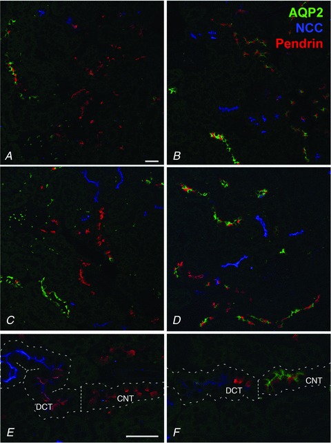
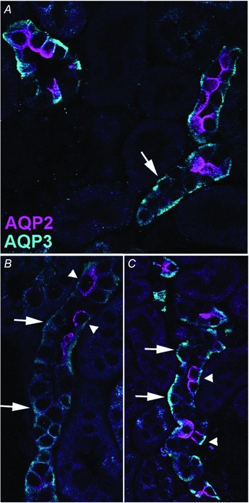
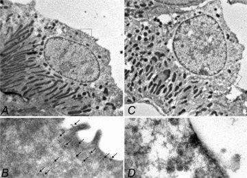
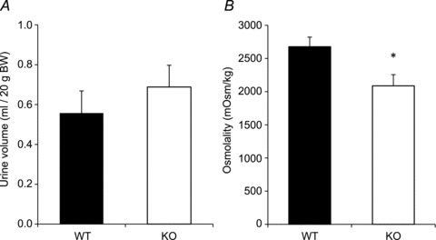
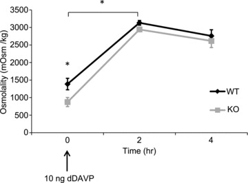
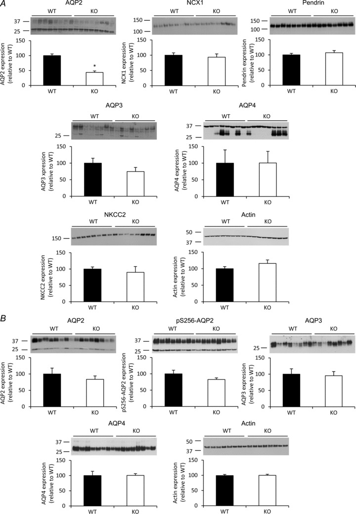
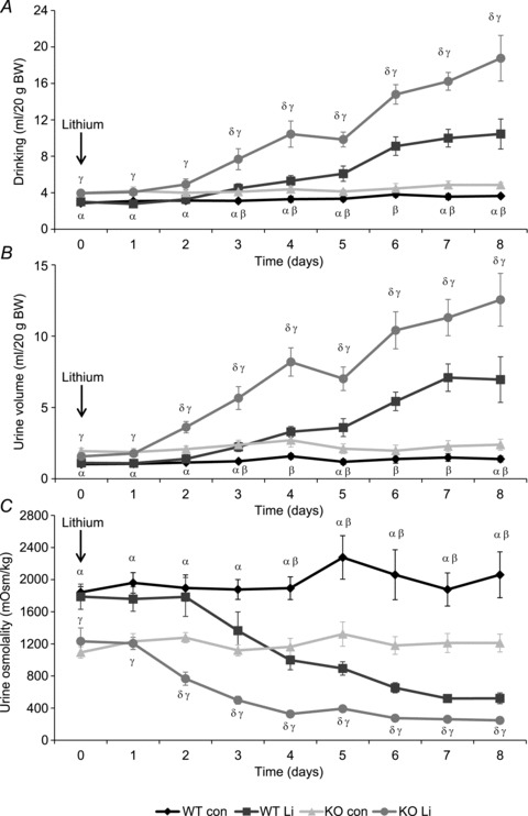
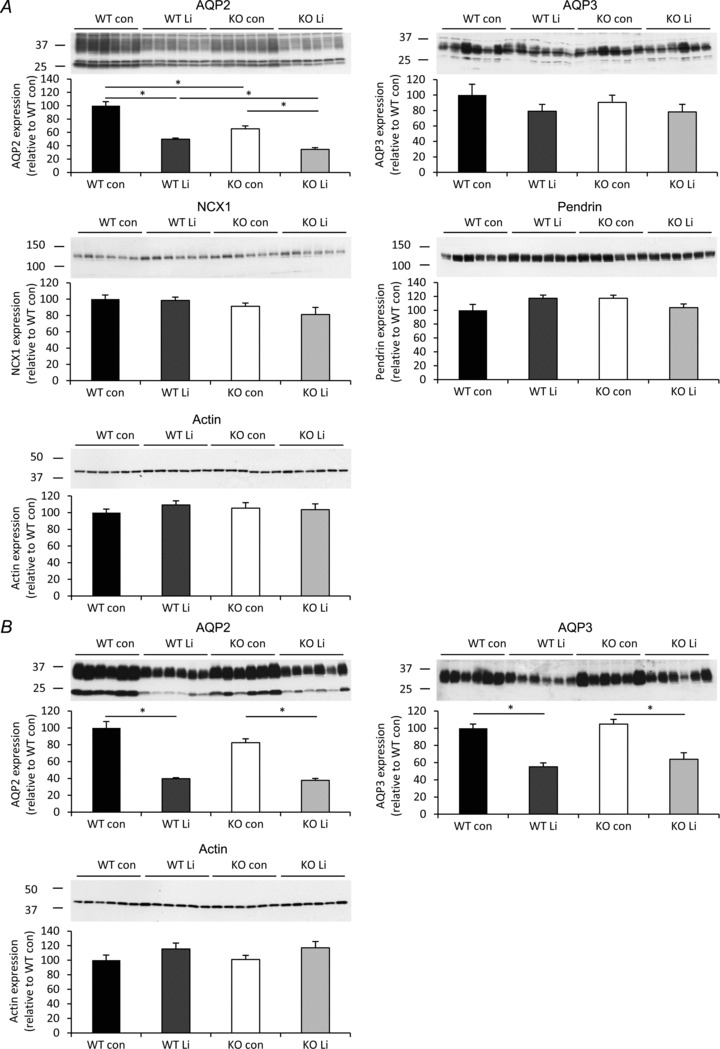
Body water balance is regulated via the water channel aquaporin-2 (AQP2), which is expressed in the renal connecting tubule (CNT) and collecting duct (CD). The relative roles of AQP2 in the CNT and CD are not fully understood. To study the role of AQP2 in the CNT we generated a mouse model with CNT-specific AQP2 deletion (AQP2-CNT-knockout (KO)). Confocal laser scanning microscopy and immunogold electron microscopy demonstrated an absence of AQP2 in the CNT of AQP2-CNT-KO mice. Twenty-four hour urine output was significantly increased (KO: 3.0 ± 0.3 ml (20 g body weight (BW))(-1); wild-type (WT): 1.9 ± 0.3 ml (20 g BW)(-1)) and urine osmolality decreased (KO: 1179 ± 107 mosmol kg(-1); WT: 1790 ± 146 mosmol kg(-1)) in AQP2-CNT-KO mice compared with controls. After 24 h water restriction, urine osmolality was still significantly lower in AQP2-CNT-KO mice (KO: 2087 ± 169 mosmol kg(-1); WT: 2678 ± 144 mosmol kg(-1)). A significant difference in urine osmolality between groups before desmopressin (dDAVP) (KO: 873 ± 129 mosmol kg(-1); WT: 1387 ± 163 mosmol kg(-1)) was not apparent 2 h after injection, with urine osmolality increased significantly in both groups (KO: 2944 ± 41 mosmol kg(-1); WT: 3133 ± 66 mosmol kg(-1)). Cortical kidney fractions from AQP2-CNT-KO mice had significantly reduced AQP2, with no compensatory changes in sodium potassium chloride cotransporter (NKCC2), AQP3 or AQP4. Lithium chloride treatment increased urine volume and decreased osmolality in both WT and AQP2-CNT-KO mice. After 8 days of treatment, the AQP2-CNT-KO mice still had a significantly higher urine volume and lower urine osmolality, suggesting that the CNT does not play a significant role in the pathology of lithium-induced nephrogenic diabetes insipidus. Our studies indicate that the CNT plays a role in regulating body water balance under basal conditions, but not for maximal concentration of the urine during antidiuresis.


Figures

Figure 1
Figure 1. Representative laser scanning confocal immunofluorescence triple labelling of kidney cortex from aquaporin-2 (AQP2)-connecting tubule (CNT)-knockout (KO) mice (A, C and E) and control mice (B, D and F)
Each image is from a different animal. Late distal convoluted tubule (DCT), CNT and early cortical collecting duct (CCD) B-type intercalated cell marker pendrin is pseudo-labelled in red; DCT marker NCC is pseudo-labelled in blue; and AQP2 is pseudo-labelled in green. AQP2-CNT-KO mice have long segments of distal tubule solely immunolabelling for pendrin in contrast to the control mice having co-labelling of pendrin and AQP2 in similar segments, demonstrating deletion of AQP2 in the CNT. E, the transition from NCC-positive DCT to pendrin-positive CNT is indicated by the dotted line, and only control mice (F) show co-labelling with AQP2 in the pendrin-positive CNT. Scale bar: 200 μm.
Figure 2
Figure 2. Representative laser scanning confocal immunofluorescence double labelling of kidney cortex from aquaporin-2 (AQP2)-connecting tubule (CNT)-knockout (KO) mice
AQP2 is pseudo-labelled in pink and AQP3 in turquoise. Large arrows highlight AQP3-positive but AQP2-negative cells, while arrowheads highlight cells positive for both AQP3 and AQP2.
Figure 3
Figure 3. Immunogold electron microscopy of connecting tubule (CNT) cells in kidney cortex from control (A, B) and aquaporin-2 (AQP2)-CNT-knockout (KO) mice (C, D)
Cells were labelled with AQP2 (15 nm gold particle) as well as with H-ATPase as a marker of intercalated cells (5 nm particle, not observed). Clear AQP2 expression was visible in CNT principal cells of control mice, whereas no labelling was observed in CNT principal cells of KO mice.
Figure 4
Figure 4. Basal 24 h urine volume (A) and urine osmolality (B) of control (wild-type (WT)) and aquaporin-2 (AQP2)-connecting tubule (CNT)-knockout (KO) mice (KO)
Values are mean ± SEM. *P < 0.05.
Figure 5
Figure 5. Twenty-four hour urine volume (A) and urine osmolality (B) of control (wild-type (WT)) and aquaporin-2 (AQP2)-connecting tubule (CNT)-knockout (KO) mice (KO) after 24 h water restriction
Values are mean ± SEM. *P < 0.05.
Figure 6
Figure 6. Urine osmolality of control (wild-type (WT)) and aquaporin-2 (AQP2)-connecting tubule (CNT)-knockout (KO) mice (KO) after intraperitoneal injection of desmopressin (dDAVP)
Values are mean ± SEM. *P < 0.05.
Figure 7
Figure 7. Immunoblots of cortical and inner medullary kidney homogenates
A, the abundance of aquaporin (AQP)2, AQP3, AQP4, sodium/calcium exchanger 1 (NCX1), pendrin, sodium potassium chloride cotransporter (NKCC2) and actin was determined in cortical kidney homogenates from either control (wild-type (WT)) or AQP2-connecting tubule (CNT)-knockout (KO) mice (KO). B, the expression of AQP2, pS256-AQP2, AQP3, AQP4 and actin was determined in inner medullary kidney homogenates from the same animals. Data are band densities relative to WT (mean ± SEM) after normalization to actin expression. n= 8 in the WT group, n= 8 in the KO group. *P < 0.05.
Figure 8
Figure 8. Effects of lithium chloride treatment on urinary concentration in aquaporin-2 (AQP2)-connecting tubule (CNT)-knockout (KO) mice
Twenty-four hour drinking volume (A), urine volume (B) and urine osmolality (C) of control (wild-type (WT)) and AQP2-CNT-KO mice (KO) during 8 days of lithium chloride diet (Li) or control diet (con). n= 6 per group. Values are mean ± SEM. αP < 0.05 WT con vs. KO con; βP < 0.05 WT con vs. WT Li; γP < 0.05 WT Li vs. KO Li; δP < 0.05 KO con vs. KO Li.
Figure 9
Figure 9. Immunoblots of cortical and inner medullary kidney homogenates of mice treated without (con) or with lithium chloride (Li) for 8 days
A, the expression of aquaporin (AQP)2, AQP3, sodium/calcium exchanger 1 (NCX1), pendrin and actin was determined in cortical kidney homogenates from either wild-type (WT) or AQP2-connecting tubule (CNT)-knockout (KO) mice (KO). B, the expression of AQP2, AQP3 and actin was determined in inner medullary kidney homogenates from the same animals. Data are band densities relative to WT (mean ± SEM) after normalization to actin expression. n= 6 in each group. *P < 0.05.

References

    1. Bailey MA, Cantone A, Yan Q, MacGregor GG, Leng Q, Amorim JB, Wang T, Hebert SC, Giebisch G, Malnic G. Maxi-K channels contribute to urinary potassium excretion in the ROMK-deficient mouse model of Type II Bartter's syndrome and in adaptation to a high-K diet. Kidney Int. 2006;70:51–59. - PubMed
    1. Boton R, Gaviria M, Batlle DC. Prevalence, pathogenesis, and treatment of renal dysfunction associated with chronic lithium therapy. Am J Kidney Dis. 1987;10:329–345. - PubMed
    1. Christensen BM, Kim YH, Kwon TH, Nielsen S. Lithium treatment induces a marked proliferation of primarily principal cells in rat kidney inner medullary collecting duct. Am J Physiol Renal Physiol. 2006;291:F39–F48. - PubMed
    1. Christensen BM, Marples D, Kim YH, Wang W, Frokiaer J, Nielsen S. Changes in cellular composition of kidney collecting duct cells in rats with lithium-induced NDI. Am J Physiol Cell Physiol. 2004;286:C952–C964. - PubMed
    1. Christensen BM, Perrier R, Wang Q, Zuber AM, Maillard M, Mordasini D, Malsure S, Ronzaud C, Stehle JC, Rossier BC, Hummler E. Sodium and potassium balance depends on alphaENaC expression in connecting tubule. J Am Soc Nephrol. 2010;21:1942–1951. - PMC - PubMed

Publication types

LinkOut - more resources