

Hepatocellular tumors: immunohistochemical analyses for classification and prognostication
- PMID: 23359751
- PMCID: PMC3551308
- DOI: 10.1007/s11670-011-0245-6
Hepatocellular tumors: immunohistochemical analyses for classification and prognostication
Abstract
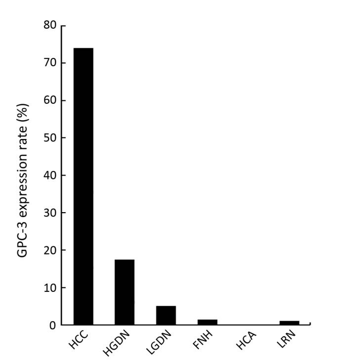
Following the classification of hepatocellular nodules by the International Working Party in 1995 and further elaboration by the International Consensus Group for Hepatocellular Neoplasia in 2009, entities under the spectrum of hepatocellular nodules have been better characterized. Research work hence has been done to answer questions such as distinguishing high-grade dysplastic nodules from early hepatocellular carcinoma (HCC), delineating the tumor cell origin of HCC, identifying its prognostic markers, and subtyping hepatocellular adenomas. As a result, a copious amount of data at immunohistochemical and molecular levels has emerged. A panel of immunohistochemical markers including glypican-3, heat shock protein 70 and glutamine synthetase has been found to be of use in the diagnosis of small, well differentiated hepatocellular tumors and particularly of HCC. The use of liver fatty acid binding protein (L-FABP), β-catenin, glutamine synthetase, serum amyloid protein and C-reactive protein is found to be helpful in the subtyping of hepatocellular adenomas. The role of tissue biomarkers for prognostication in HCC and the use of biomarkers in subclassifying HCC based on tumor cell origin are also discussed.
Keywords: Classification; Hepatocellular tumors; Immunohistochemical.
Figures
References
-
- Wanless IR. Terminology of nodular hepatocellular lesions. Hepatology 1995;22:983-93 - PubMed
-
- Kojiro M, Roskams T.Early hepatocellular carcinoma and dysplastic nodules. Semin Liver Dis 2005;25:133-42 - PubMed
-
- Roskams T, Kojiro M.Pathology of early hepatocellular carcinoma: conventional and molecular diagnosis. Semin Liver Dis 2010;30:17-25 - PubMed
-
- Kojiro M, Wanless IR, Alves V, et al. Pathologic diagnosis of early hepatocellular carcinoma: A report of the international consensus group for hepatocellular neoplasia. Hepatology 2009;49:658-64 - PubMed
-
- Bioulac-Sage P, Balabaud C, Zucman-Rossi J.Subtype classifi- cation of hepatocellular adenoma. Dig Surg 2010;27:39-45 - PubMed
LinkOut - more resources
Full Text Sources
Research Materials